
上浒充通大学 Shanghai Jiao Tong University Lecture 3-1: Microbial cell biology Chapter 3 in 8 BROCK BIOLOGY OF MICROORGANISMS AIJIAO TONG UNI Zhao Liping,Chen Feng School of Life Science and Technology, Shanghai Jiao Tong University http://micro.sjtu.edu.cn
Lecture 3-1: Microbial cell biology Chapter 3 in BROCK BIOLOGY OF MICROORGANISMS Zhao Liping, Chen Feng School of Life Science and Technology, Shanghai Jiao Tong University http://micro.sjtu.edu.cn

上商充通大学 Shanghai Jiao Tong University I.Microscopy and cell morphology 3.1 Light Microscopy 3.2 Three-Dimentsional Imaging:Interference Contrast, Atomic Force,and Confocal Scanning 3.3 Electron Mciroscopy 3.4 Cell Morphology and the Significance of Being Small Shanghai Jiao Tong University
Shanghai Jiao Tong University I. Microscopy and cell morphology 3.1 Light Microscopy 3.2 Three-Dimentsional Imaging: Interference Contrast, Atomic Force, and Confocal Scanning 3.3 Electron Mciroscopy 3.4 Cell Morphology and the Significance of Being Small
上商充至大学 Shanghai Jiao Tong University 3.1 Light Microscopy SHANGHAL JIAO TONG UN mnvn 光学显微镜
3.1 Light Microscopy 光学显微镜
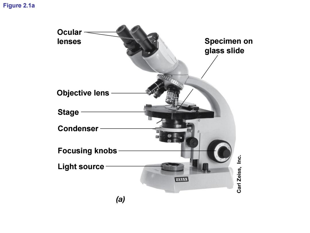
Figure 2.1a Ocular lenses Specimen on glass slide Objective lens Stage Condenser Focusing knobs Light source ZEISS a
Figure 2.1a Ocular lenses Objective lens Stage Condenser Focusing knobs Light source Specimen on glass slide
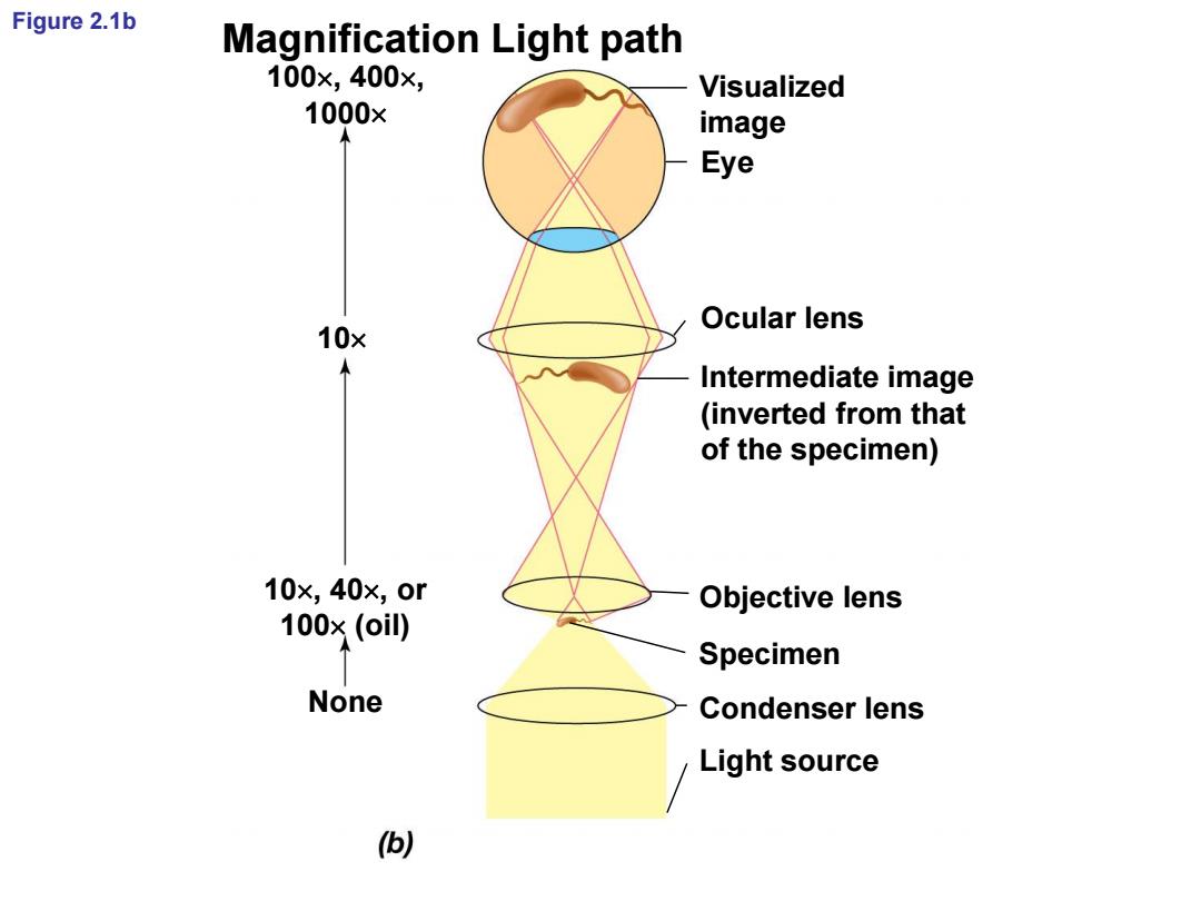
Figure 2.1b Magnification Light path 100×,400×, Visualized 1000× image Eye Ocular lens 10× Intermediate image (inverted from that of the specimen) 10x,40x,or Objective lens 100×(oil) Specimen None Condenser lens Light source )
Figure 2.1b Visualized image Eye Ocular lens Intermediate image (inverted from that of the specimen) Objective lens Specimen Condenser lens Light source None 100, 400, 1000 10 10, 40, or 100 (oil) Magnification Light path
上泽充豆大学 Shanghai Jiao Tong University Staining:Increasing Contrast for Bright-Field Microscopy SHA 8 染色:增加明视野显微镜的对比度 AUIAO TONG UNA
Staining: Increasing Contrast for Bright-Field Microscopy 染色: 增加明视野显微镜的对比度

上泽充豆大学 Shanghai Jiao Tong University Staining:Increasing Contrast for Bright-Field Microscopy Staining:Increasing Contrast for Bright-Field Microscopy Staining:Increasing Contrast for Bright-Field Microscopy Shanghai Jiao Tong University
Shanghai Jiao Tong University Staining: Increasing Contrast for Bright-Field Microscopy Staining: Increasing Contrast for Bright-Field Microscopy Staining: Increasing Contrast for Bright-Field Microscopy
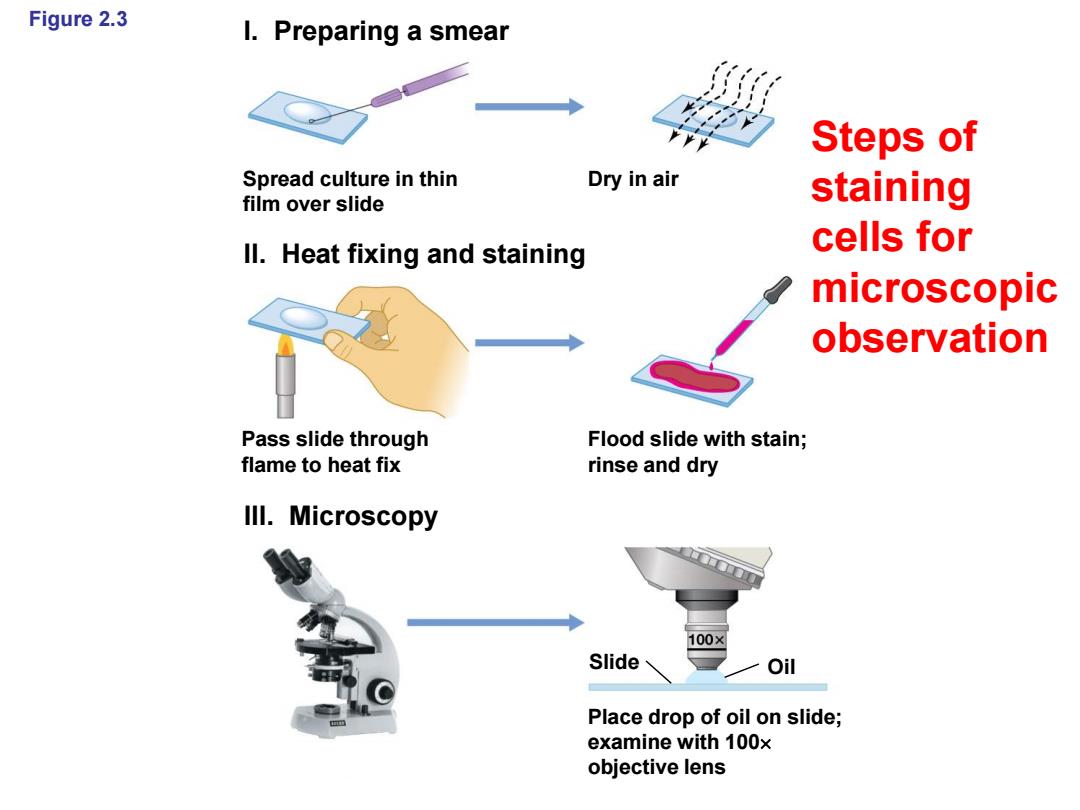
Figure 2.3 I.Preparing a smear Steps of Spread culture in thin Dry in air film over slide staining Il.Heat fixing and staining cells for microscopic observation Pass slide through Flood slide with stain; flame to heat fix rinse and dry Ill.Microscopy 100× Slide Oil Place drop of oil on slide; examine with 100x objective lens
Figure 2.3 I. Preparing a smear II. Heat fixing and staining III. Microscopy Spread culture in thin film over slide Pass slide through flame to heat fix Dry in air Flood slide with stain; rinse and dry Place drop of oil on slide; examine with 100 objective lens Slide Oil Steps of staining cells for microscopic observation
上商充通大学 Shanghai Jiao Tong University T Differential Stains:The Gram Stain SHANG 鉴定染色:革兰氏染色法 AIJIAO TONG UNI
Differential Stains: The Gram Stain 鉴定染色: 革兰氏染色法
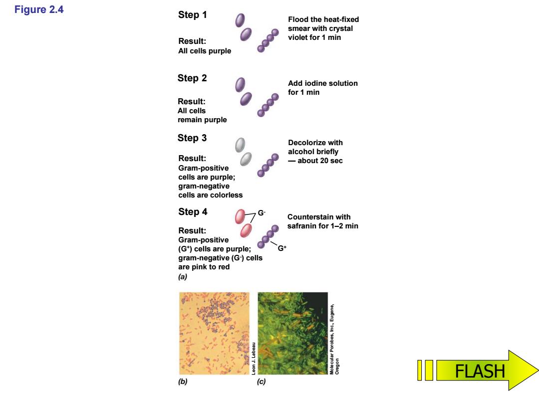
Figure 2.4 Step 1 Flood the heat-fixed smear with crystal Result: violet for 1 min All cells purple Step 2 Add iodine solution for 1 min Result: All cells remain purple Step 3 Decolorize with alcohol briefly Result: 一about20sec Gram-positive cells are purple; gram-negative cells are colorless Step4 Counterstain with Result: safranin for 1-2 min Gram-positive (G)cells are purple; gram-negative(G-)cells are pink to red (a) FLASH (c)
Figure 2.4 Step 1 Step 2 Step 3 Step 4 Result: All cells purple Result: All cells remain purple Result: Gram -positive cells are purple; gram -negative cells are colorless Result: Gram -positive (G + ) cells are purple; gram -negative (G - ) cells are pink to red Flood the heat -fixed smear with crystal violet for 1 min Add iodine solution for 1 min Decolorize with alcohol briefly — about 20 sec Counterstain with safranin for 1 –2 min G + G - Leon J. Lebeau Molecular Porobes, Inc., Eugene, Oregon FLASH