Myeloma proteins If we are going to study the structure of antibodies they must be purified and antibodies are difficult to purify since they have the same chemical and physical properties.One way around the purification problem is to use myeloma proteins which can be obtained in pure form from multiple myeloma patients. Plasma cells normally form one specific kind of antibody in relatively small quantities.It was observed that plasma cells can be transformed into tumor plasma cells,called myeloma cells.The transformed cells produce monoclonal antibodies of a single type since each tumor consist of identical cells.The antibodies may be in the form of an intact globulin,light chain peptide or heavy chain peptide. s of cells which 4C entibody released dentical Antibody(IgG)
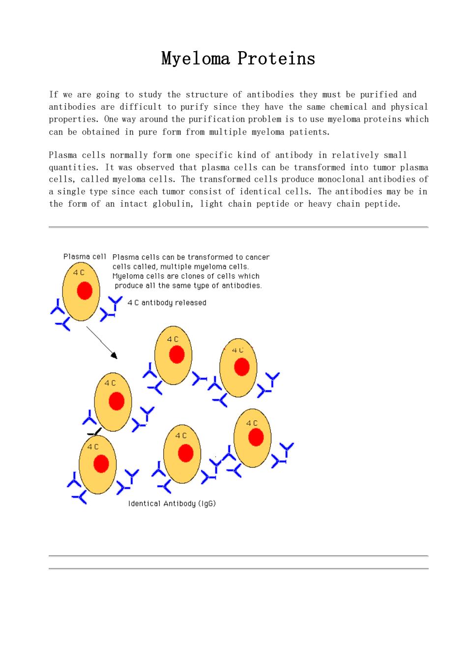
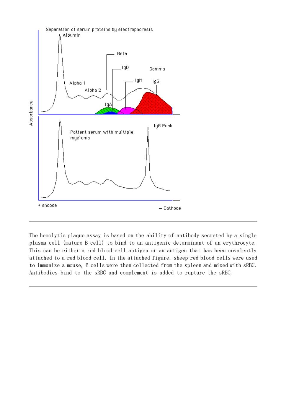
Myeloma Proteins If we are going to study the structure of antibodies they must be purified and antibodies are difficult to purify since they have the same chemical and physical properties. One way around the purification problem is to use myeloma proteins which can be obtained in pure form from multiple myeloma patients. Plasma cells normally form one specific kind of antibody in relatively small quantities. It was observed that plasma cells can be transformed into tumor plasma cells, called myeloma cells. The transformed cells produce monoclonal antibodies of a single type since each tumor consist of identical cells. The antibodies may be in the form of an intact globulin, light chain peptide or heavy chain peptide

S6parntinmomanmproteinstyolectroptorosie Beta IgG Peak Patient serum with multiple myeloma andode -Cethode The hemolytic plaque assay is based on the ability of antibody secreted by a single plasma cell (mature B cell)to bind to an antigenic determinant of an erythrocyte. This can be either a red blood cell antigen or an antigen that has been covalently sheep red blood cells were used Antibodies bind to the sRBC and complement is added to rupture the sRBC
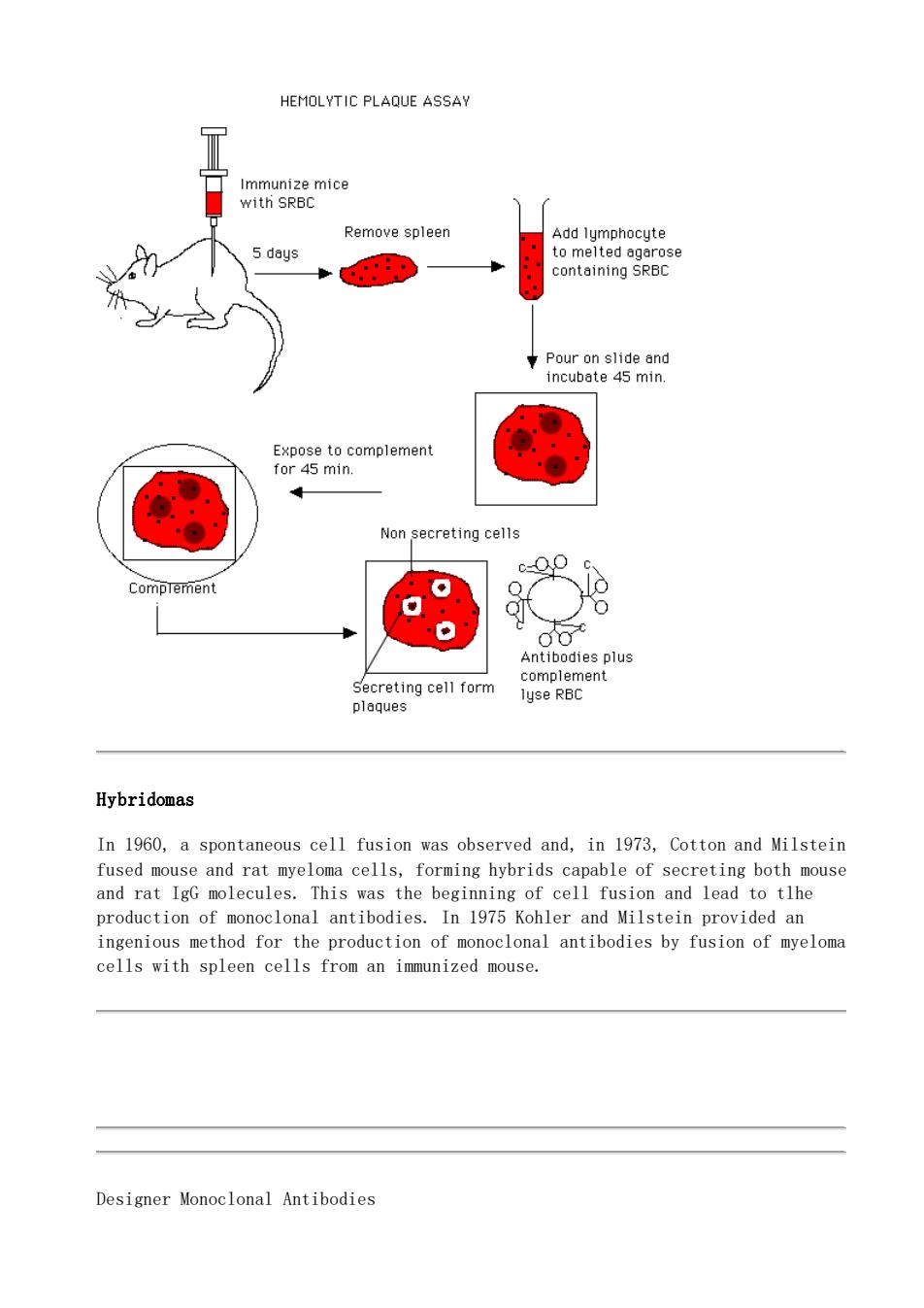
The hemolytic plaque assay is based on the ability of antibody secreted by a single plasma cell (mature B cell) to bind to an antigenic determinant of an erythrocyte. This can be either a red blood cell antigen or an antigen that has been covalently attached to a red blood cell. In the attached figure, sheep red blood cells were used to immunize a mouse, B cells were then collected from the spleen and mixed with sRBC. Antibodies bind to the sRBC and complement is added to rupture the sRBC

HEMOLYTIC PLAQUE ASSAY Remove spleer Add lymphocyte 5 days Pour on slide and incubate 45 min. Non secreting cells CompTement 9 Antibodies plus Hybridomas In 1960,a spontaneous cell fusion was observed and,in 1973,Cotton and Milstein and rat IgG molecules ingenious method for the production of monoclonal antibodies by fusion of myeloma cells with spleen cells from an immunized mouse. Designer Monoclonal Antibodies
Hybridomas In 1960, a spontaneous cell fusion was observed and, in 1973, Cotton and Milstein fused mouse and rat myeloma cells, forming hybrids capable of secreting both mouse and rat IgG molecules. This was the beginning of cell fusion and lead to tlhe production of monoclonal antibodies. In 1975 Kohler and Milstein provided an ingenious method for the production of monoclonal antibodies by fusion of myeloma cells with spleen cells from an immunized mouse. Designer Monoclonal Antibodies

Designer Monoclonal Antibodies mice After Five Days emove Spleen ◆ CDNA Restriction ..CCA Enzymes Digestion 品 and Ligated Digestion Inserted Restriction Enzymes 124 Transfer to E.coli Detect clones by an antigen Ahi6ooging The mRNA is isolated from B cells in the spleen and used to produce complementary (DNA).The DNA sequence for Ig,H and L chains,is amplified by polymerase chain reaction (PCR)using primers.The H and L chain gene products are then digested with restriction enzymes into small DNA fragments.These are then inserted(ligated)into viral DNA of a phage and the phage is used to infect E.coli.The virus grows lytically and antibody chains can be detected with antigens. Return
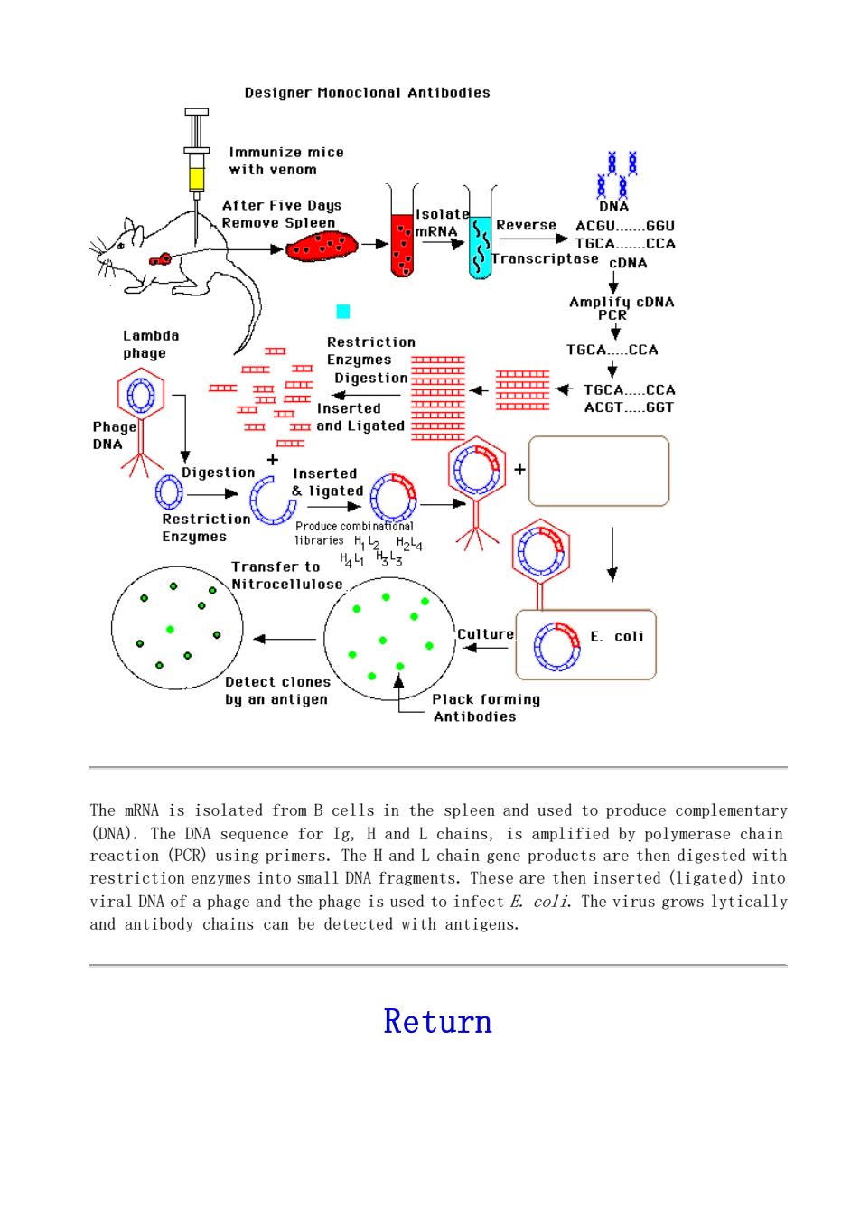
The mRNA is isolated from B cells in the spleen and used to produce complementary (DNA). The DNA sequence for Ig, H and L chains, is amplified by polymerase chain reaction (PCR) using primers. The H and L chain gene products are then digested with restriction enzymes into small DNA fragments. These are then inserted (ligated) into viral DNA of a phage and the phage is used to infect E. coli. The virus grows lytically and antibody chains can be detected with antigens. Return

This page is maintained by the Natural Toxins Research Center at Texas A&M University -Kingsville
This page is maintained by the Natural Toxins Research Center at Texas A&M University - Kingsville