Introduction to Techniques in Immunology Introduction to the Immune System Immunology emerged from medical science and has permeated all biology.The spread of immunology into other fields has been the result of the scientist and clinician using immunologic techniques as a sensitive analytical tool. Immunological assays are important in regulatory work because of their sensitivity,specificity,and rapidity.To aid in understanding the theory behind immunoassays a brief introduction to the immune system will be presented. We live in a hostile world filled with many infectious agents;however vertebrate animals possess an effective immune system which prevents the invasions of parasites such as bacteria,viruses,and cancer cells.The immune system specifically recognizes and selectively eliminates foreign invaders.The immune systems responds in a specific way to pathogens and displays a long-term memory of earlier contacts with the disease agents. Vertebrates are protected by a dual immune system known as cell-mediated immunity and humoral immunity.The two immune systems together provide an excellent defense against foreign invaders.Both systems are adaptive and respond specificallytomost foreign substances,although depending on the antigen one immune response generally is favored over the other. Cell-mediated immunity is particularly effective against fungi,parasites intracellular viral infections,cancer cells,and foreign tissue.The
Introduction to Techniques in Immunology Introduction to the Immune System Immunology emerged from medical science and has permeated all biology. The spread of immunology into other fields has been the result of the scientist and clinician using immunologic techniques as a sensitive analytical tool. Immunological assays are important in regulatory work because of their sensitivity, specificity, and rapidity. To aid in understanding the theory behind immunoassays a brief introduction to the immune system will be presented. We live in a hostile world filled with many infectious agents; however, vertebrate animals possess an effective immune system which prevents the invasions of parasites such as bacteria, viruses, and cancer cells. The immune system specifically recognizes and selectively eliminates foreign invaders. The immune systems responds in a specific way to pathogens and displays a long-term memory of earlier contacts with the disease agents. Vertebrates are protected by a dual immune system known as cell-mediated immunity and humoral immunity. The two immune systems together provide an excellent defense against foreign invaders. Both systems are adaptive and respond specifically to most foreign substances, although depending on the antigen one immune response generally is favored over the other. Cell-mediated immunity is particularly effective against fungi, parasites, intracellular viral infections, cancer cells, and foreign tissue. The
humoral immune response defends primarily against extracellular bacteria and viral infections.The cells involved in both immune systems are lymphocytes which originate in the bone marrow and migrate to different lymphoid organs.There are two types of lymphocytes which are known as T cells and B cells.The two types of lymphocytes are responsible for a dual immunity phases Figure 1.All lymphocytes are derived from bone marrow but those that pass through the thymus become T cells.(T lymphocytes).The T cells are responsible for cell-mediated immunity.Since the immunity involving T cells is associated with the T cells themselves,this type of immunity is called cell-mediated immunity. Other lymphocytes pass through the bursa of Fabricius in birds or the bursa equivalent in mammals and become B cells (B lymphocytes).The bursa of Fabricius is the primary lymphoid organ associated with the cloaca in birds but is not found in mammals.The bursa-derived lymphocytes(B cells)produce antibodies which can react specifically with antigen.Because B cells produce antibodies that circulate and the immunity is called humoral immunity.Antigen specific antibodies produced in the humoral immune response can be used in immunological assays to detect various disease agents or antigenic molecules.With the development of monoclonal antibodies,greater specificity and reproducibility can be obtained with immunological assays.The remainder of this discussion will be devoted to the humoral immune response. The Humoral Immune Response
humoral immune response defends primarily against extracellular bacteria and viral infections. The cells involved in both immune systems are lymphocytes which originate in the bone marrow and migrate to different lymphoid organs. There are two types of lymphocytes which are known as T cells and B cells. The two types of lymphocytes are responsible for a dual immunity phases Figure 1. All lymphocytes are derived from bone marrow but those that pass through the thymus become T cells. (T lymphocytes). The T cells are responsible for cell-mediated immunity. Since the immunity involving T cells is associated with the T cells themselves, this type of immunity is called cell-mediated immunity. Other lymphocytes pass through the bursa of Fabricius in birds or the bursa equivalent in mammals and become B cells (B lymphocytes). The bursa of Fabricius is the primary lymphoid organ associated with the cloaca in birds but is not found in mammals. The bursa-derived lymphocytes (B cells) produce antibodies which can react specifically with antigen. Because B cells produce antibodies that circulate and the immunity is called humoral immunity. Antigen specific antibodies produced in the humoral immune response can be used in immunological assays to detect various disease agents or antigenic molecules. With the development of monoclonal antibodies, greater specificity and reproducibility can be obtained with immunological assays. The remainder of this discussion will be devoted to the humoral immune response. The Humoral Immune Response
Basic to the humoral immune response is the formation of antibodies (protein molecules)generated in response to the presence of foreign substances. The foreign substances that induce an immune response and interact with antibodies are called antigens (or immunogens).Antigens are traditionally defined as any substance that,when introduced parenterally into an animal, will cause the production of antibodies and will react specifically with the antibodies in vitro (Figure 2).Antigens are macromolecules (10,000 MW)that possess a high degree of internal chemical complexity.They are soluble in water and foreign to the animal in which they stimulate antibodies (Figure 3&4).We generally do not produce antibodies against our own body's molecules or against low-molecular-weight molecules (less than 10,000). Haptens are low molecular weight molecules that are non-antigenic and cannot stimulate antibody production by themselves;however,they will react with appropriate antibody molecules.When haptens combinewitha larger carrier molecule,they convey a new antigenic determinant site to the carrier molecule.Drugs and pesticides are low molecular weight molecules and can be treated as haptens phases Figure 5.By conjugation to larger carrier molecules (albumin),low molecular weight drugs and pesticides can be made antigenic.Antibodies produced in this matter will react specifically with low molecular weight haptens. The following are characteristics of antigens
Basic to the humoral immune response is the formation of antibodies (protein molecules) generated in response to the presence of foreign substances. The foreign substances that induce an immune response and interact with antibodies are called antigens (or immunogens). Antigens are traditionally defined as any substance that, when introduced parenterally into an animal, will cause the production of antibodies and will react specifically with the antibodies in vitro (Figure 2). Antigens are macromolecules (10,000 MW) that possess a high degree of internal chemical complexity. They are soluble in water and foreign to the animal in which they stimulate antibodies (Figure 3 & 4). We generally do not produce antibodies against our own body's molecules or against low-molecular-weight molecules (less than 10,000). Haptens are low molecular weight molecules that are non-antigenic and cannot stimulate antibody production by themselves; however, they will react with appropriate antibody molecules. When haptens combine with a larger carrier molecule, they convey a new antigenic determinant site to the carrier molecule. Drugs and pesticides are low molecular weight molecules and can be treated as haptens phases Figure 5. By conjugation to larger carrier molecules (albumin), low molecular weight drugs and pesticides can be made antigenic. Antibodies produced in this matter will react specifically with low molecular weight haptens. The following are characteristics of antigens
Molecular size A good antigen is a macromolecule that has molecular weight of 10,000 or more.Proteins like insulin Figure 2 Figure 3 and 4 Figure 4. (5,700 mol wt)are poor antigens.Polysaccharides like heparin (17,000 mol wt)are also nonantigenic and do not produce antibodies when injected into animals.The greater the molecular weight of a substance,the more likely it is to function as an antigen.Within each molecule there are specific regions of limited size (10,000 mol wt)that function as the antigenic determinant sites.Larger antigens will have more antigenic determinant sites which means that many different antibodies will be produced in response to a large antigen Molecular complexity High molecular weight is not enough to confer antigenicity on a foreign substance.There must be internal complexity The organic chemist can produce synthetic polymers of any size but most are not antigenic unless there is internal complexity.Most naturally occurring macromolecules are often very complex because they are built from many different low molecular weight constituents.Proteins are very antigenic since they consist of 20 different amino acids. Solubility Another argument used to explain the nonantigenicity of synthetic polymers is their insolubility in body fluids.Many of the particulate antigens,bacteria and viruses are engulfed by macrophages and digested into soluble components
Molecular size A good antigen is a macromolecule that has molecular weight of 10,000 or more. Proteins like insulin Figure 2 Figure 3 and 4 Figure 4. (5,700 mol wt) are poor antigens. Polysaccharides like heparin (17,000 mol wt) are also nonantigenic and do not produce antibodies when injected into animals. The greater the molecular weight of a substance, the more likely it is to function as an antigen. Within each molecule there are specific regions of limited size (10,000 mol wt) that function as the antigenic determinant sites. Larger antigens will have more antigenic determinant sites which means that many different antibodies will be produced in response to a large antigen. Molecular complexity High molecular weight is not enough to confer antigenicity on a foreign substance. There must be internal complexity. The organic chemist can produce synthetic polymers of any size but most are not antigenic unless there is internal complexity. Most naturally occurring macromolecules are often very complex because they are built from many different low molecular weight constituents. Proteins are very antigenic since they consist of 20 different amino acids. Solubility Another argument used to explain the nonantigenicity of synthetic polymers is their insolubility in body fluids. Many of the particulate antigens, bacteria and viruses are engulfed by macrophages and digested into soluble components
Foreignness Antigens must be foreign to the host.The more foreign the antigen is to the host,the better it will stimulate antibody formation. Duck serum proteins are not good antigens for chickens,but antigen from bacteria are extremely antigenic and will stimulate the formation of antibodies in chickens.Animals do not generally produce antibodies against self protein. When a vertebrate first encounters an antigen,it exhibits a primary humoral immune response.If the animal encounters the same antigen after a few days the immune resonse is more rapid and has a greater magnitude (Figure 6) The initial encounter causes specific B-cell clones to proliferate and differentiate.The progeny lymphocytes include not only effector cells (antibody producing cells)but also clones of memory cells,which retain the capacity to produce both effector and memory cells upon subsequent stimulation by the original antigen.The effector cells live for only a few days;therefore,the antibody titer increases and decreases within 20 days.The memory cells live for a lifetime and can be reactivated by a second stimuation with the same antigen.Thus when an antigen is encountered a second time,its memory cells quickly produce effector cells which rapidly produce massive quantities of antibodies.The secondary immune response is also called the anamnestic response or booster response. Figure 5 Figure 6 Figure 7
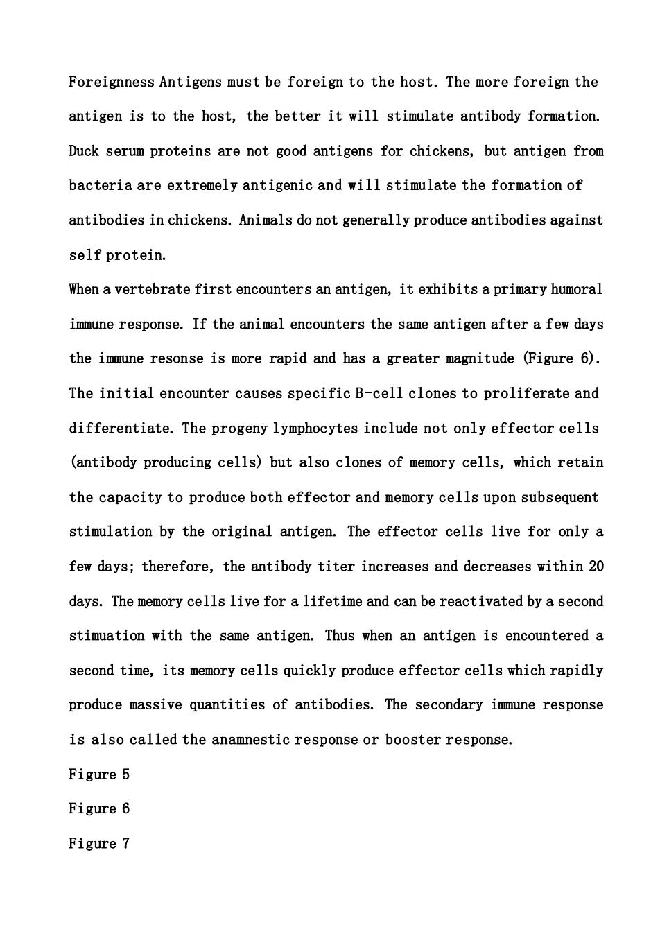
Foreignness Antigens must be foreign to the host. The more foreign the antigen is to the host, the better it will stimulate antibody formation. Duck serum proteins are not good antigens for chickens, but antigen from bacteria are extremely antigenic and will stimulate the formation of antibodies in chickens. Animals do not generally produce antibodies against self protein. When a vertebrate first encounters an antigen, it exhibits a primary humoral immune response. If the animal encounters the same antigen after a few days the immune resonse is more rapid and has a greater magnitude (Figure 6). The initial encounter causes specific B-cell clones to proliferate and differentiate. The progeny lymphocytes include not only effector cells (antibody producing cells) but also clones of memory cells, which retain the capacity to produce both effector and memory cells upon subsequent stimulation by the original antigen. The effector cells live for only a few days; therefore, the antibody titer increases and decreases within 20 days. The memory cells live for a lifetime and can be reactivated by a second stimuation with the same antigen. Thus when an antigen is encountered a second time, its memory cells quickly produce effector cells which rapidly produce massive quantities of antibodies. The secondary immune response is also called the anamnestic response or booster response. Figure 5 Figure 6 Figure 7
Antibodies Antibodies are Y-shaped proteins found in sera which are produced in response to a specific antigen(Figure 7).Antibodies have different molecular weights and sedimentation coefficients depending on the class of antibodies.IgG has a molecular weight of 150,000 and a sedimentation coefficient of 7S.The largest antibody molecule is IgM with a molecular weight of 900,000 and a sedimentation coefficient of 19S. Antibodies are composed of two heavy peptide chains and two short peptide chains (Figure 7).Two identical heavy chains have a molecular weight of 50,000 each,and two identical light chains have a molecular weight of 25,000 each.These chains are connected by inter-disulfide bonds.Purified preparations of IgG are resistant to reductive cleavage by sulfhydryl reagents unless the molecule was unfolded by high concentrations of urea or guanidine.The number and precise position of both inter and intra disulfide bonds differ and are a characteristic of the subclasses.At the amino terminal end of the antibody is a short segment called the variable region.The amino acid sequence of the variable region is different for each antibody and is specific for a certain antigen.Within each variable region there is a hypervariable region.The hypervariable region of the antibody binds specifically to the antigen in a lock-and-key manner.It is the variable region of the antibody which allows development of sensitive and specific immunoassays. The carboxyl end of the heavy and light chain of the antibody molecule is called the constant region.The amino acid sequence of the constant region
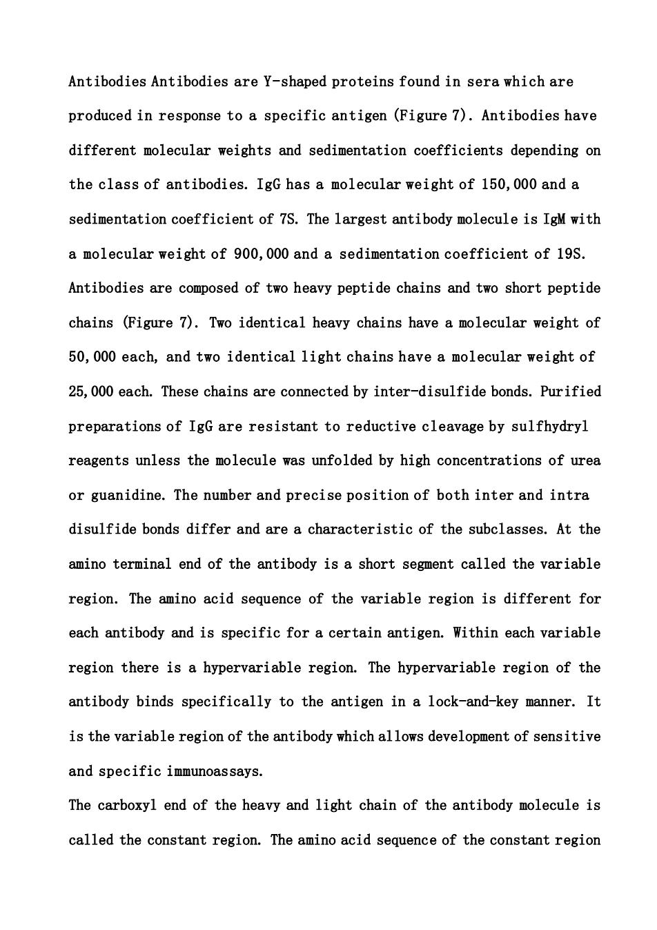
Antibodies Antibodies are Y-shaped proteins found in sera which are produced in response to a specific antigen (Figure 7). Antibodies have different molecular weights and sedimentation coefficients depending on the class of antibodies. IgG has a molecular weight of 150,000 and a sedimentation coefficient of 7S. The largest antibody molecule is IgM with a molecular weight of 900,000 and a sedimentation coefficient of 19S. Antibodies are composed of two heavy peptide chains and two short peptide chains (Figure 7). Two identical heavy chains have a molecular weight of 50,000 each, and two identical light chains have a molecular weight of 25,000 each. These chains are connected by inter-disulfide bonds. Purified preparations of IgG are resistant to reductive cleavage by sulfhydryl reagents unless the molecule was unfolded by high concentrations of urea or guanidine. The number and precise position of both inter and intra disulfide bonds differ and are a characteristic of the subclasses. At the amino terminal end of the antibody is a short segment called the variable region. The amino acid sequence of the variable region is different for each antibody and is specific for a certain antigen. Within each variable region there is a hypervariable region. The hypervariable region of the antibody binds specifically to the antigen in a lock-and-key manner. It is the variable region of the antibody which allows development of sensitive and specific immunoassays. The carboxyl end of the heavy and light chain of the antibody molecule is called the constant region. The amino acid sequence of the constant region

is similar to the sequence of antibody in the same class.The constant region is the part of the antibody that binds to mast cells,complement,and protein A.Protein A is a bacterial cell wall protein isolated from Staphylococcus aureus which binds to the Fc (Fragment-crystallizable)fragment of the antibody molecule.This protein can be used in affinity chromatography to purify antibodies. Papain digestion cleaves IgG molecules into two fragments which can be separated by carboxymethyl cellulose ion exchange chromatography.One fragment will crystallize spontaneously.This fragment is called Fe fragment (fragment-crystallizable)and is deficient in antigen-binding ability.The Fc fragment has a molecular weight of approximately 50,000 and a sedimentation coefficient of 3.5S.The Fc fragment is the constant region of the heavy chain with a COOH terminus end.The second fragment is fragment antigen-binding (Fab).The molecular weight and sedimentation coefficient for the Fab are similar to Fc fragments.An IgG molecule consists of 1 Fc and 2 Fab fragments.Each Fab fragment consists of the amino terminal half of the heavy and light chains.It was possible to predict the structure of IgG molecule with Papain (Fc and Fab)fragments and sulfhydryl reagents which split the IgG molecule into light and heavy chains Antibodies specifically combine with a small segment of the antigen called the antigenic determinant or epitope to form an antigen-antibody complex. The antigen-antibody reaction is characterized by specificity.If a mouse is injected with goat albumin,the mouse will produce antigoat albumin
is similar to the sequence of antibody in the same class. The constant region is the part of the antibody that binds to mast cells, complement, and protein A. Protein A is a bacterial cell wall protein isolated from Staphylococcus aureus which binds to the Fc (Fragment-crystallizable) fragment of the antibody molecule. This protein can be used in affinity chromatography to purify antibodies. Papain digestion cleaves IgG molecules into two fragments which can be separated by carboxymethyl cellulose ion exchange chromatography. One fragment will crystallize spontaneously. This fragment is called Fc fragment (fragment-crystallizable) and is deficient in antigen-binding ability. The Fc fragment has a molecular weight of approximately 50,000 and a sedimentation coefficient of 3.5S. The Fc fragment is the constant region of the heavy chain with a COOH terminus end. The second fragment is fragment antigen-binding (Fab). The molecular weight and sedimentation coefficient for the Fab are similar to Fc fragments. An IgG molecule consists of 1 Fc and 2 Fab fragments. Each Fab fragment consists of the amino terminal half of the heavy and light chains. It was possible to predict the structure of IgG molecule with Papain (Fc and Fab) fragments and sulfhydryl reagents which split the IgG molecule into light and heavy chains. Antibodies specifically combine with a small segment of the antigen called the antigenic determinant or epitope to form an antigen-antibody complex. The antigen-antibody reaction is characterized by specificity. If a mouse is injected with goat albumin, the mouse will produce antigoat albumin
antibodies.These specific antibodies will react only with goat albumin or albumin that is closely related (Figure 2).This process is referred to as a polyclonal response.The polyclonal response is partly due to the different clones of B cells of the immune system and partly due to the complex nature of antigens.The polyclonal response suggests that different clones of B cells are producing five different classes of antibodies to various epitopes which have diverse affinities.When a disease agent attacks a vertebrate host,a whole army of different antibodies are effective in neutralizing the disease agent (Figure 8).These same polyclonal antibodies can be used in immunoassays;however,the wide-variety of antibodies make the assay less specific.With the development of monoclonal antibodies greater specificity can be achieved in most immunoassays. Types of antigen-antibody reactions Basically there are two types of serological reactions.Agglutination reactions occur between an antibody and particulate antigens such as RBC or bacteria (Figure 9).Agglutination of bacteria has been a key method in identifying and classifying microorganisms. Precipitation reactions occur between an antibody and a fluid antigen. Precipitation reactions are most useful since most antigens are soluble or can be solubilized by simple procedures.In addition,there are many types of precipitation test which can be adapted to many types of antigens
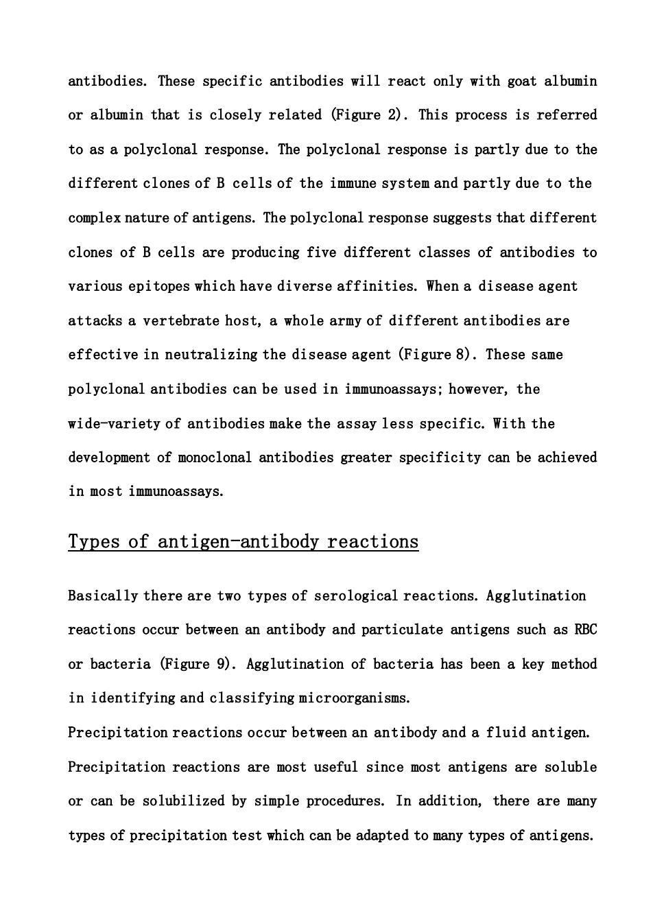
antibodies. These specific antibodies will react only with goat albumin or albumin that is closely related (Figure 2). This process is referred to as a polyclonal response. The polyclonal response is partly due to the different clones of B cells of the immune system and partly due to the complex nature of antigens. The polyclonal response suggests that different clones of B cells are producing five different classes of antibodies to various epitopes which have diverse affinities. When a disease agent attacks a vertebrate host, a whole army of different antibodies are effective in neutralizing the disease agent (Figure 8). These same polyclonal antibodies can be used in immunoassays; however, the wide-variety of antibodies make the assay less specific. With the development of monoclonal antibodies greater specificity can be achieved in most immunoassays. Types of antigen-antibody reactions Basically there are two types of serological reactions. Agglutination reactions occur between an antibody and particulate antigens such as RBC or bacteria (Figure 9). Agglutination of bacteria has been a key method in identifying and classifying microorganisms. Precipitation reactions occur between an antibody and a fluid antigen. Precipitation reactions are most useful since most antigens are soluble or can be solubilized by simple procedures. In addition, there are many types of precipitation test which can be adapted to many types of antigens
In many cases nonprecipitating antigen-antibody reactions can be detected indirectly with other antibodies,isotopes,or enzymes. The antigen-antibody reaction takes place in two phases Figure 10.In the first phase,combination of the reactants occurs:this is followed by second phase,an aggregation (precipitation or agglutination).The first phase stages take place almost instantaneously.In the second stage of the reaction,aggregation of the antigen-antibody complex occurs.This phase does not occur when monovalent antibodies or haptens are involved in the reaction.The lattice hypothesis,which is based on multivalent antigens and bivalent antibodies,is useful in explaining the zone phenomena.At the equivalence point all of the antigen and all the antibody molecules are consumed in the lattice formation.With multivalent antigen where Figure 8 Figure 9 the antigenic site is not repeated,it requires two different antibodies directed at different antigenic sites to form a lattice.When there is an excess of antibody (prozone),aggregation is not observed phases Figure 11.Each antigen is surround by antibodies.Essentially the reverse situation occurs in postzone,where too little antibody is present to produce an aggregation each antibody molecule only binds to one antigen. The size of the aggregation complex increases as the optimal ratio of antigen to antibody is achieved.This is referred to as the equivalence zone
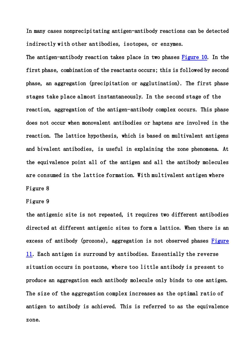
In many cases nonprecipitating antigen-antibody reactions can be detected indirectly with other antibodies, isotopes, or enzymes. The antigen-antibody reaction takes place in two phases Figure 10. In the first phase, combination of the reactants occurs; this is followed by second phase, an aggregation (precipitation or agglutination). The first phase stages take place almost instantaneously. In the second stage of the reaction, aggregation of the antigen-antibody complex occurs. This phase does not occur when monovalent antibodies or haptens are involved in the reaction. The lattice hypothesis, which is based on multivalent antigens and bivalent antibodies, is useful in explaining the zone phenomena. At the equivalence point all of the antigen and all the antibody molecules are consumed in the lattice formation. With multivalent antigen where Figure 8 Figure 9 the antigenic site is not repeated, it requires two different antibodies directed at different antigenic sites to form a lattice. When there is an excess of antibody (prozone), aggregation is not observed phases Figure 11. Each antigen is surround by antibodies. Essentially the reverse situation occurs in postzone, where too little antibody is present to produce an aggregation each antibody molecule only binds to one antigen. The size of the aggregation complex increases as the optimal ratio of antigen to antibody is achieved. This is referred to as the equivalence zone
There are many immunological assays based upon optimal precipitation of an antigen and antibody.A few of the most commonly used methods to characterize monoclonals and soluble antigens are two-dimensional immunoelectrophoresis,rocket electrophoresis,countercurrent electrophoresis,radial immuno-diffusion,immunoelectrophoresis,and Western blot.Each of these are briefly explained in Figure 12,Figure 13, Figure 14,Figure 15 figures 12-16. Monoclonal Antibody Production. A monoclonal antibody is produced by a single clone of B cells that has a single specificity and is of one class or subclass.The production of monoclonal antibodies begins with the injection of a mouse with a particular antigen phases Figure 17. A major advantage of this procedure is that the antigen used need not be purified,as is the case in polyclonal procedures for producing a specific antiserum.In a few weeks,when the B lymphocytes begin proliferating in response to the antigen,the spleen,a primary source of these lymphocytes, is removed from the mouse.See figure 17 for details of the production of monoclonal antibodies.Among the many lymphocytes isolated from this mouse spleen are a few B cells which produce the desired antibody.These spleen cells are fused in vitro with myeloma cells,a special tumor lymphocyte that multiplies unchecked.Like most tumor cells,these myeloma cells can
There are many immunological assays based upon optimal precipitation of an antigen and antibody. A few of the most commonly used methods to characterize monoclonals and soluble antigens are two-dimensional immunoelectrophoresis, rocket electrophoresis, countercurrent electrophoresis, radial immuno-diffusion, immunoelectrophoresis, and Western blot. Each of these are briefly explained in Figure 12, Figure 13, Figure 14, Figure 15 figures 12-16. Monoclonal Antibody Production. A monoclonal antibody is produced by a single clone of B cells that has a single specificity and is of one class or subclass. The production of monoclonal antibodies begins with the injection of a mouse with a particular antigen phases Figure 17. A major advantage of this procedure is that the antigen used need not be purified, as is the case in polyclonal procedures for producing a specific antiserum. In a few weeks, when the B lymphocytes begin proliferating in response to the antigen, the spleen, a primary source of these lymphocytes, is removed from the mouse. See figure 17 for details of the production of monoclonal antibodies. Among the many lymphocytes isolated from this mouse spleen are a few B cells which produce the desired antibody. These spleen cells are fused in vitro with myeloma cells, a special tumor lymphocyte that multiplies unchecked. Like most tumor cells, these myeloma cells can