Disorders of the orbit
Disorders of the orbit

Anatomy of the orbit The orbit refers to the bony cavity in the skull FRONTAL BONE SPHENOID Supra-orbital that houses the eye and surrounding structures. notch Optic canal Diseases of the orbit can arise from within the ETHMOID orbit or as part of a systemic illness that affects LACRIMAL multiple tissues or organs. Inferior orbital nssure PALATINE 日ONE infra-orbital infra-orbital MAXILLA Some signs oforbital disorders include: groove foramen Protrusion of the eyeball ·Pain Diplopia or double vision ·Loss of vision Redness and swelling of the eyelids
Anatomy of the orbit • The orbit refers to the bony cavity in the skull that houses the eye and surrounding structures. Diseases of the orbit can arise from within the orbit or as part of a systemic illness that affects multiple tissues or organs. • Some signs of orbital disorders include: • • Protrusion of the eyeball • Pain • Diplopia or double vision • Loss of vision • Redness and swelling of the eyelids

Orbital diseases 。Thyroid eye disease Orbital infection and inflammations ·Orbital tumors Congenital orbital malformations
Orbital diseases • Thyroid eye disease • Orbital infection and inflammations • Orbital tumors • Congenital orbital malformations
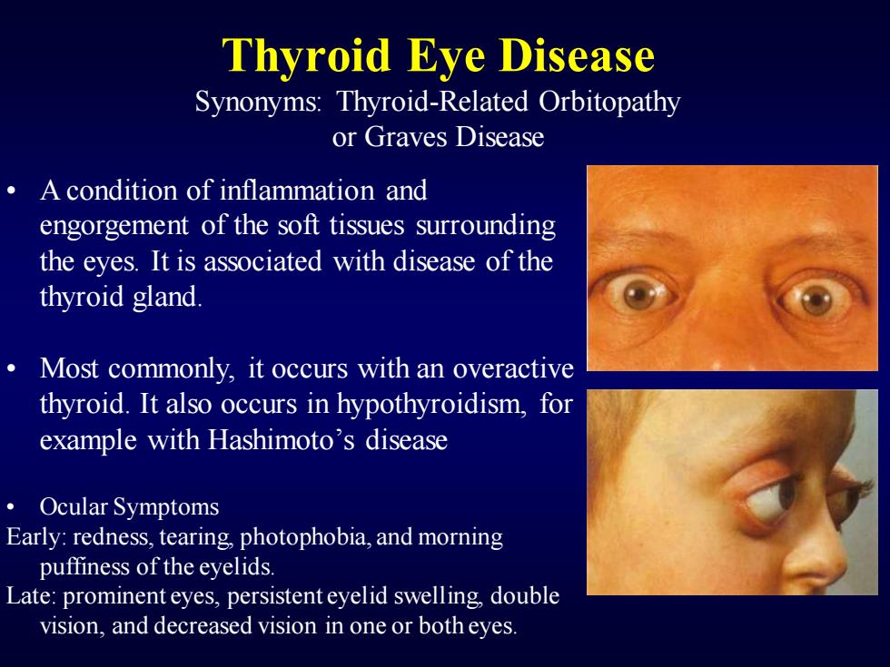
Thyroid Eye Disease Synonyms:Thyroid-Related Orbitopathy or Graves Disease A condition of inflammation and engorgement of the soft tissues surrounding the eyes.It is associated with disease of the thyroid gland. Most commonly,it occurs with an overactive thyroid.It also occurs in hypothyroidism,for example with Hashimoto's disease ·Ocular Symptoms Early:redness,tearing,photophobia,and morning puffiness of the eyelids. Late:prominent eyes,persistenteyelid swelling,double vision,and decreased vision in one or both eyes
Thyroid Eye Disease Synonyms: Thyroid-Related Orbitopathy or Graves Disease • A condition of inflammation and engorgement of the soft tissues surrounding the eyes. It is associated with disease of the thyroid gland. • Most commonly, it occurs with an overactive thyroid. It also occurs in hypothyroidism, for example with Hashimoto’s disease • Ocular Symptoms Early: redness, tearing, photophobia, and morning puffiness of the eyelids. Late: prominent eyes, persistent eyelid swelling, double vision, and decreased vision in one or both eyes
Pathology Activated T cells infiltrate orbital contents and stimulate fibroblasts,leading to: 1.Enlargement of extraocular muscles 2.Cellular infiltration of interstitial tissues 3.Proliferation of orbital fat and connective tissue
Pathology Activated T cells infiltrate orbital contents and stimulate fibroblasts, leading to: 1.Enlargement of extraocular muscles 2.Cellular infiltration of interstitial tissues 3.Proliferation of orbital fat and connective tissue

Enlargement of extraocular muscles The stimulated fibroblasts produce glycosaminoglycans (GAGs)which cause the muscle to swell Muscle size may increase by up Swollen muscles to 8 times The swollen muscles occupy orbital space and can compress Compression the optic nerve ofoptic nerve These swollen muscles can at apex oforbit cause a forward propulsion of the globe (proptosis)so that the eyelids do not cover well and Swollen muscle(medialrectus) eyes dry out,causing exposure keratopathy
Enlargement of extraocular muscles • The stimulated fibroblasts produce glycosaminoglycans (GAGs) which cause the muscle to swell • Muscle size may increase by up to 8 times • The swollen muscles occupy orbital space and can compress the optic nerve • These swollen muscles can cause a forward propulsion of the globe (proptosis) so that the eyelids do not cover well and eyes dry out, causing exposure keratopathy Swollen muscles Compression of optic nerve at apex of orbit Swollen muscle (medial rectus)
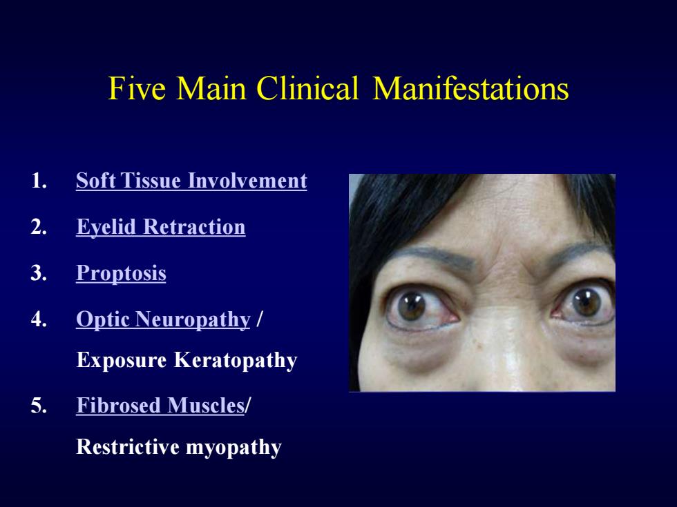
Five Main Clinical Manifestations 1. Soft Tissue Involvement 2. Eyelid Retraction 3. Proptosis 4. Optic Neuropathy Exposure Keratopathy 5. Fibrosed Muscles/ Restrictive myopathy
Five Main Clinical Manifestations 1. Soft Tissue Involvement 2. Eyelid Retraction 3. Proptosis 4. Optic Neuropathy / Exposure Keratopathy 5. Fibrosed Muscles/ Restrictive myopathy
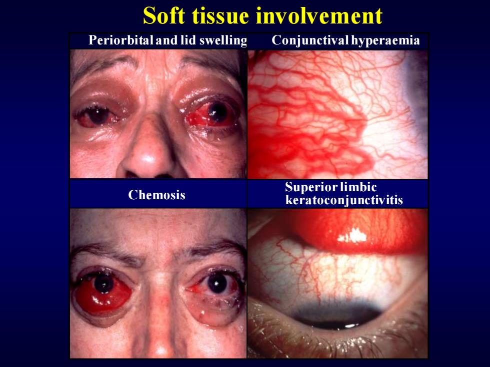
Soft tissue involvement Periorbitaland lid swelling Conjunctival hyperaemia Superior limbic Chemosis keratoconjunctivitis
Soft tissue involvement Periorbital and lid swelling Chemosis Conjunctival hyperaemia Superior limbic keratoconjunctivitis

Signs of eyelid retraction Occurs in about 50% Bilaterallid retraction Bilaterallid retraction No associated proptosis.Bilateral proptosis Unilateral lid retraction ·Lid lag in downgaze Unilateral proptosis
Signs of eyelid retraction Occurs in about 50% • Bilateral lid retraction • No associated proptosis • Bilateral lid retraction • Bilateral proptosis • Lid lag in downgaze • Unilateral lid retraction • Unilateral proptosis

Proptosis Occurs in about 50% Uninfluenced by treatment of hyperthyroidism Axial and permanent in about 70% May be associated with choroidal folds Treatment options ·Systemic steroids ·Radiotherapy Surgical decompression
Proptosis Treatment options • Systemic steroids • Radiotherapy • Surgical decompression • Occurs in about 50% • Uninfluenced by treatment of hyperthyroidism Axial and permanent in about 70% May be associated with choroidal folds