Week 16 Quiz 2 Female reproductivepathology 1.A32-year-od woman has experienced ull pelvic pain for the past month Physical examination showsa right adnexal mass.An abdominal ultrasound scan shows a 7.5-cm cystic ovarian mass.The mass is surgically excised.The surface of the mass is smooth,and it is not adherent to surrounding pelvic structures.On gross examination the mass is cystic and filled with hair.Microscopically,there is a squamous epithelium,tall columnar glandular epithelium,cartilage,and fibrous connective tissue present.Which of the following is the MOST LIKELY diagnosis? A.Adenocarcinoma B.Fibroadenoma C.Mesothelioma D.Rhabdomyosarcoma E Teratoma AnsE suface.They are not inasive and therefore case e nd in Cha ciive age neesy seb they are mesoderm onnective tissue o'a】ysmo'e prope ystadenoc cinomas presen clinically with lower abdominal pain and abdominal enlargement.Gl complaints,urinary frequency,dysuria and pelvic pressure may occur.Pelvic adhesions due to tumor infiltration and metastasis at the time of diagnosis are common.Tumors are characteristically solid and poorly differentiated.Fibroadenoma is a common tumor that occurs in the breast.Fibroma is a benign,solid tumor of the ovary.Mesothelioma was commonly arises in the pleural cavity.It may rarely occur in the peritoneum.It is a diffusely infiltrating tumor that causes symptoms by causing ascites or bowel obstruction.Rhabdomyosarcoma is rare tumor which occurs in the vagina of female infants and more commonly in the head.neck and extremities of children less than 20 vears. 2.A 35-year-old woman had a firm nodule palpable on the dome of the uterus six years ago recorded on routine physical examination.The nodule has slowly increased in size and is now appears to be about twice the size it was when first discovered.She remains asymptomatic.Which of the following neoplasms is she MOST LIKELY to have? A.Adenocarcinoma B.Hemangioma Leiomyoma omyosarcoma Ans:C Leiomyomas,commonly called fibroids.are perhaps the most common tumor in women.They are benign smooth muscle neoplasms that occur singly but are commonly multiple.Leiomyomas are commonly asymptomatic even if they are large and multiple.They are hormonally responsive but grow very slowly. Adenocarcinoma of the uterus arises from the endometrium.it comes to medical attention because it produces irregular or postmenopausal bleeding.Uterine enlargement may be absent.It does not produce a firm palpable nodule.Hemangioma is a benign tumor of childhood.It is most common in the skin and mucous membranes. Hemangiomas are also commonly seen in the liver of adults.Leiomyosarcoma is uncommon.There is a peak incidence at 40-60 years of age.They grow rapidly.Ovarian carcinomas generally metastasize to the
Week 16 Quiz 2 Female reproductivepathology 1. A 32-year-old woman has experienced dull pelvic pain for the past 2 months. Physical examination shows a right adnexal mass. An abdominal ultrasound scan shows a 7.5-cm cystic ovarian mass. The mass is surgically excised. The surface of the mass is smooth, and it is not adherent to surrounding pelvic structures. On gross examination the mass is cystic and filled with hair. Microscopically, there is a squamous epithelium, tall columnar glandular epithelium, cartilage, and fibrous connective tissue present. Which of the following is the MOST LIKELY diagnosis? A. Adenocarcinoma B. Fibroadenoma C. Mesothelioma D. Rhabdomyosarcoma E. Teratoma Ans: E Mature teratomas are benign tumors with a smooth surface. They are not invasive and therefore cause no adhesion to adjacent structures. Teratomas are usually found in young women of reproductive age. Characteristically, they are cystic and contain hair and cheesy sebaceous material. Histologically, they are composed of tissues derived from epiderm (epithelium) and mesoderm (bone, cartilage and connective tissue). Adenocarcinoma of the ovary is more properly termed Cystadenocarcinoma. Cystadenocarcinomas present clinically with lower abdominal pain and abdominal enlargement. GI complaints, urinary frequency, dysuria and pelvic pressure may occur. Pelvic adhesions due to tumor infiltration and metastasis at the time of diagnosis are common. Tumors are characteristically solid and poorly differentiated. Fibroadenoma is a common tumor that occurs in the breast. Fibroma is a benign, solid tumor of the ovary. Mesothelioma was commonly arises in the pleural cavity. It may rarely occur in the peritoneum. It is a diffusely infiltrating tumor that causes symptoms by causing ascites or bowel obstruction. Rhabdomyosarcoma is rare tumor which occurs in the vagina of female infants and more commonly in the head, neck and extremities of children less than 20 years. 2. A 35-year-old woman had a firm nodule palpable on the dome of the uterus six years ago recorded on routine physical examination. The nodule has slowly increased in size and is now appears to be about twice the size it was when first discovered. She remains asymptomatic. Which of the following neoplasms is she MOST LIKELY to have? A. Adenocarcinoma B. Hemangioma C. Leiomyoma D. Leiomyosarcoma E. Metastastic ovarian carcinoma Ans: C Leiomyomas, commonly called fibroids, are perhaps the most common tumor in women. They are benign smooth muscle neoplasms that occur singly but are commonly multiple. Leiomyomas are commonly asymptomatic even if they are large and multiple. They are hormonally responsive but grow very slowly. Adenocarcinoma of the uterus arises from the endometrium. It comes to medical attention because it produces irregular or postmenopausal bleeding. Uterine enlargement may be absent. It does not produce a firm palpable nodule. Hemangioma is a benign tumor of childhood. It is most common in the skin and mucous membranes. Hemangiomas are also commonly seen in the liver of adults. Leiomyosarcoma is uncommon. There is a peak incidence at 40-60 years of age. They grow rapidly. Ovarian carcinomas generally metastasize to the

Detoem and catse ascltes They suall tmole the uerine sijace bin more commonby present as d infiltrative process rather that a slow growing mass eold woman has noricedaody discharge from the nipple of her right be amination,th cycle A.Acute mastitis B.Fibroadenoma C.Intraductal papilloma D.Phylloides tumor E.Sclerosingadenosis Ans:C Intraductalpapillomas are usually solitary and smaller than i cm.They are located in large lactiferous sinuses or ducts and have a tendency to bleed.Acute mastitis occurs most commonly in lactating women and is painful. It does not produce bloody dischars e.Fibroadenomas contain ducts with stroma and are not highly vascular Theyre odPhireromratobl nalignant.They ade ducts to cause bleeding.Sclerosingadenosi s.a lesion occurring with fibrocystic changes.has abundant collagen but not vascularity. 4.A 44-year-old woman sees her physician because she felt a lump in her left breast I week ago.The physician palpates a firm,irregular mass in the upper outer quadrant of the left breast.There are no overlying skin lesions. The mammogram is shown in A and the gross appearance of the excisional biopsy specimen is shown in B above.Which of the following additional findings is MOST LIKELY to be present on physical examination? A.Axillary lymphadenopathy B.Bloody discharge from the nipple C.Cushingoid face
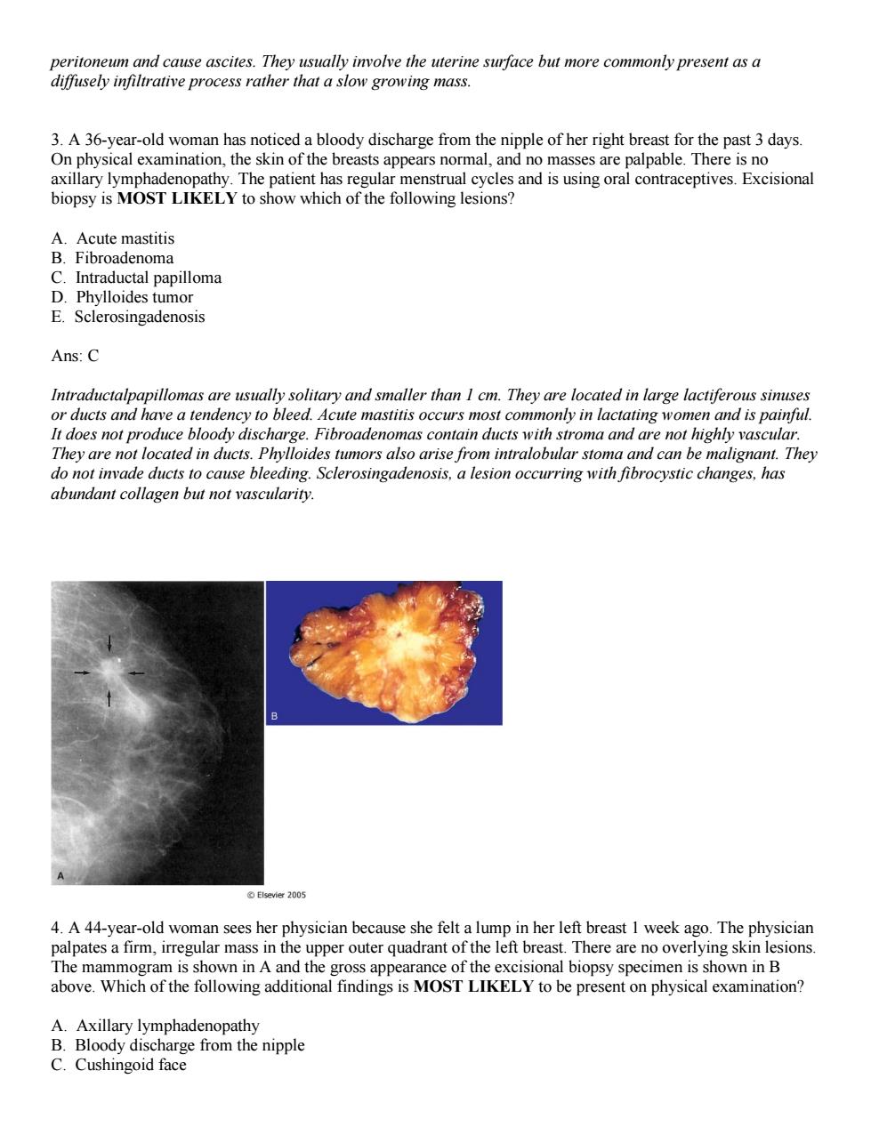
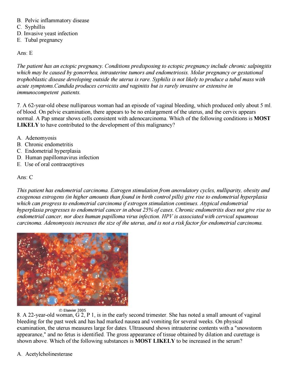
peritoneum and cause ascites. They usually involve the uterine surface but more commonly present as a diffusely infiltrative process rather that a slow growing mass. 3. A 36-year-old woman has noticed a bloody discharge from the nipple of her right breast for the past 3 days. On physical examination, the skin of the breasts appears normal, and no masses are palpable. There is no axillary lymphadenopathy. The patient has regular menstrual cycles and is using oral contraceptives. Excisional biopsy is MOST LIKELY to show which of the following lesions? A. Acute mastitis B. Fibroadenoma C. Intraductal papilloma D. Phylloides tumor E. Sclerosingadenosis Ans: C Intraductalpapillomas are usually solitary and smaller than 1 cm. They are located in large lactiferous sinuses or ducts and have a tendency to bleed. Acute mastitis occurs most commonly in lactating women and is painful. It does not produce bloody discharge. Fibroadenomas contain ducts with stroma and are not highly vascular. They are not located in ducts. Phylloides tumors also arise from intralobular stoma and can be malignant. They do not invade ducts to cause bleeding. Sclerosingadenosis, a lesion occurring with fibrocystic changes, has abundant collagen but not vascularity. 4. A 44-year-old woman sees her physician because she felt a lump in her left breast 1 week ago. The physician palpates a firm, irregular mass in the upper outer quadrant of the left breast. There are no overlying skin lesions. The mammogram is shown in A and the gross appearance of the excisional biopsy specimen is shown in B above. Which of the following additional findings is MOST LIKELY to be present on physical examination? A. Axillary lymphadenopathy B. Bloody discharge from the nipple C. Cushingoid face

Ans:A This irregular,infiltrative mass is an infiltrating ductal carcinoma.the most common form of breast cancer. Breast carcinomas are most likely to metastasize to regional lymph nodes.By the time a breast cancer becomes palpable,lymph node metastasis are present in more than 50%of patients.A bloody discharge from the nipple most often results from an intraductal papilloma.Breast cancers are associated in rare cases with ectopic corticotropin secretion or Cushing syndrome.Lobular carcinomas are more often bilateral,but they are less common than infiltrating ductal carcinomas.Pain with breast enlargement suggests inflammation. 5.A 47-year-old woman has noticed a red,scaly area of skin on her left breast that has grown slightly larger over the past 4 months.On physical examination,there is a 1-cm area of eczematous skin just lateral to the earance of the skin biopsy specimen is shown above.Which of the following is the metaplasia E.Paget disease of the breast Ans:E Paget cells are large cells that have clear mucinous cytoplasm and infiltrate the skin.They are malignant and extend to the skin from an underlying breast carcinoma.Apocrine metaplasia affects cells lining the cystically dilated ducts in fibrocystic change.The macrophages in fat necrosis do not infiltrate the skin and do not have the atypical nuclei seen in the figure."Inflammatory carcinoma"does not refer to a specific histologic type of breast cancer;rather it describes the involvement of dermal lymphatics by infiltrating carcinoma.In lobular carcinoma in situ,terminal ducts or acini are filled with neoplastic cells.The overlying skin is unaffected. 6.A 24-year-old woman experiences sudden onset of severe lower abdominal pain.Physical examination shows no masses,but there is severe tenderness in the right lower quadrant.A pelvice examination shows no lesions of the cervix or va vel sounds are detected.An abdominal ultr d shows a 4-cm focal enlar the cht fallop A dilatio dure A.Molar pregnancy
D. Mass in the opposite breast E. Painful breast enlargement Ans: A This irregular, infiltrative mass is an infiltrating ductal carcinoma, the most common form of breast cancer. Breast carcinomas are most likely to metastasize to regional lymph nodes. By the time a breast cancer becomes palpable, lymph node metastasis are present in more than 50% of patients. A bloody discharge from the nipple most often results from an intraductal papilloma. Breast cancers are associated in rare cases with ectopic corticotropin secretion or Cushing syndrome. Lobular carcinomas are more often bilateral, but they are less common than infiltrating ductal carcinomas. Pain with breast enlargement suggests inflammation. 5. A 47-year-old woman has noticed a red, scaly area of skin on her left breast that has grown slightly larger over the past 4 months. On physical examination, there is a 1-cm area of eczematous skin just lateral to the areola. The microscopic appearance of the skin biopsy specimen is shown above. Which of the following is the MOST LIKELY diagnosis? A. Apocrine metaplasia B. Fat necrosis C. Inflammatory carcinoma D. Lobular carcinoma in situ E. Paget disease of the breast Ans: E Paget cells are large cells that have clear mucinous cytoplasm and infiltrate the skin. They are malignant and extend to the skin from an underlying breast carcinoma. Apocrine metaplasia affects cells lining the cystically dilated ducts in fibrocystic change. The macrophages in fat necrosis do not infiltrate the skin and do not have the atypical nuclei seen in the figure. “Inflammatory carcinoma” does not refer to a specific histologic type of breast cancer; rather it describes the involvement of dermal lymphatics by infiltrating carcinoma. In lobular carcinoma in situ, terminal ducts or acini are filled with neoplastic cells. The overlying skin is unaffected. 6. A 24-year-old woman experiences sudden onset of severe lower abdominal pain. Physical examination shows no masses, but there is severe tenderness in the right lower quadrant. A pelvic examination shows no lesions of the cervix or vagina. Bowel sounds are detected. An abdominal ultrasound shows a 4-cm focal enlargement of the proximal right fallopian tube. A dilation and curettage procedure shows only decidua from the endometrial cavity. Which of the following is the MOST LIKELY diagnosis? A. Molar pregnancy

B.Pelvic inflammatory disease sive yeast infection E.Tubal pregnancy Ans:E The patient has an ectopic pregnancy.Conditions predisposing to ectopic pregnancy inchude chronic sapingitis which may be caused by gonorrhea,intrauterine tumors and endometriosis.Molar pregnancy or gestationa trophoblastic disease developing outside the uterus is rare.Syphilis is not likely to produce a tubal mass with acute symptoms.Candida produces cervicitis and vaginitis but is rarely invasive or extensive in immunocompetent patients. 7.A 62-year-old obese nulliparous woman had an episode of vaginal bleeding,which produced only about 5 ml of blood.On pelvic examination,there appears to be no enlargement of the uterus,and the cervix appears normal.A Pap smear shows cells consistent with adenocarcinoma.Which of the following conditions is MOST LIKELY to have contributed to the development of this malignancy? A Adenomvosis B.Chronic endometritis Endometrial hyper plasia omavirus infectior E.Use oforal contraceptives Ans:C This patient has endometrial carcinoma. Estrogen stimulation from anovulatory cycles,nulliparity.obesity and exogenous estrogens (in higher amounts than found in birth control pills)give rise to endometrial hyperplasia which can progress to endometrial carcinoma ifestrogen stimulation continues.Atypical endometrial hyperplasia progresses to endometrial cancer in about 25%of cases.Chronic endometritis does not give rise to endometrial cancer,nor does human papilloma virus infection.HPV is associated with cervical squamous carcinoma.Adenomyosis increases the size of the uterus,and is not a risk factor for endometrial carcinoma 005 8.A 22-year-old woman,G2,P 1,is in the early second trimester.She has noted a small amount of vaginal bleeding for the past week and has had marked nausea and vomiting for several weeks.On physical examination,the uterus measures large for dates.Ultrasound shows intrauterine contents with a"snowstorm appearance,"and no fetus is identified.The gross appearance of tissue obtained by dilation and curettage is shown above.Which of the following substances is MOST LIKELY to be increased in the serum? A.Acetylcholinesterase
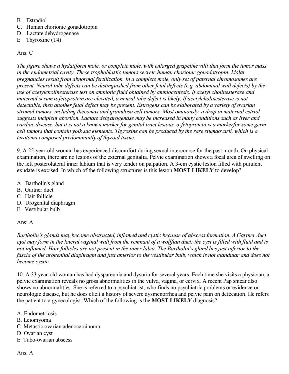
B. Pelvic inflammatory disease C. Syphillis D. Invasive yeast infection E. Tubal pregnancy Ans: E The patient has an ectopic pregnancy. Conditions predisposing to ectopic pregnancy include chronic salpingitis which may be caused by gonorrhea, intrauterine tumors and endometriosis. Molar pregnancy or gestational trophoblastic disease developing outside the uterus is rare. Syphilis is not likely to produce a tubal mass with acute symptoms.Candida produces cervicitis and vaginitis but is rarely invasive or extensive in immunocompetent patients. 7. A 62-year-old obese nulliparous woman had an episode of vaginal bleeding, which produced only about 5 ml. of blood. On pelvic examination, there appears to be no enlargement of the uterus, and the cervix appears normal. A Pap smear shows cells consistent with adenocarcinoma. Which of the following conditions is MOST LIKELY to have contributed to the development of this malignancy? A. Adenomyosis B. Chronic endometritis C. Endometrial hyperplasia D. Human papillomavirus infection E. Use of oral contraceptives Ans: C This patient has endometrial carcinoma. Estrogen stimulation from anovulatory cycles, nulliparity, obesity and exogenous estrogens (in higher amounts than found in birth control pills) give rise to endometrial hyperplasia which can progress to endometrial carcinoma if estrogen stimulation continues. Atypical endometrial hyperplasia progresses to endometrial cancer in about 25% of cases. Chronic endometritis does not give rise to endometrial cancer, nor does human papilloma virus infection. HPV is associated with cervical squamous carcinoma. Adenomyosis increases the size of the uterus, and is not a risk factor for endometrial carcinoma. 8. A 22-year-old woman, G 2, P 1, is in the early second trimester. She has noted a small amount of vaginal bleeding for the past week and has had marked nausea and vomiting for several weeks. On physical examination, the uterus measures large for dates. Ultrasound shows intrauterine contents with a "snowstorm appearance," and no fetus is identified. The gross appearance of tissue obtained by dilation and curettage is shown above. Which of the following substances is MOST LIKELY to be increased in the serum? A. Acetylcholinesterase

B.Estradiol Lactate genase E Thyroxine(T4) Ans:C The figure shows a hydatiform mole.or complete mole,with enlarged grapelike villi that form the tumor mass in the endometrial cavity.These trophoblastic tumors secrete human chorionic gonadotropin.Molar pregnancies result from abnormal fertilization.In a complete mole,only set of paternal chromosomes are present.Neural tube defects can be distinguished from other fetal defects (e.g.abdominal wall defects)by the use of acetylcholinesterase test on amniotic fluid obtained by amniocentesis.If acetyl cholinesterase and maternal serum a-fetoprotein are elevated.a neural tube defect is likely.If acetylcholinesterase is not detectable.then another fetal defect may be present.Estrogens can be elaborated by a variety of ovarian stromal tumors,including thecomas and gramulosa cell tumors.Most ominously.a drop in maternal estriol pient abortion.Lactate dehvdr nay he increased in m ns such as liver and se,but it is ot a knoy ital tract lesions nors that c protein isam ne can be produ ed by the teratoma composed pre minantly of thyroid tissu 9.A 25-year- ysical xamination,there a ons o1 examination show area of swe ng or the left posterolateral inner labium that is very tender on palpation.A 3-cm cystic lesion filled with purulent exudate is excised.In which of the following structures is this lesion MOST LIKELY to develop? A.Bartholin's gland B.Gartner duct C.Hair follicle D.Urogenital diaphragm E.Vestibular bulb Ans A Bartholin's glands may become obstructed,inflamed and cystic because of abscess formation.A Gartner duct cyst may form in the lateral caginal wall fro the ren ant of a wolffian duct:the cyst is filled with fluid and is not inflamed.Hair follicles not pre. ent in the inne The B tholin's gland lies rogenal diaphragm and just anterior to the which i s not gla 10.A 33 year-old womar has had dyspareuniaand dysuria for several years.Each time she visits a physician,a pelvic exa ent Pap smear also shows no al inthe who tinds no psychiatrie problems or evidence or to a psychiatrist, neurologic disease,but he does elicit a history or severe dysmenorrnea an d pelvic pain on defecation.He refers the patient to a gynecologist.Which of the following is the MOST LIKELY diagnosis? A.Endometriosis B.Leiomyoma C.Metastic ovarian adenocarcinoma D.Ovarian cyst E.Tubo-ovarian abscess Ans:A
B. Estradiol C. Human chorionic gonadotropin D. Lactate dehydrogenase E. Thyroxine (T4) Ans: C The figure shows a hydatiform mole, or complete mole, with enlarged grapelike villi that form the tumor mass in the endometrial cavity. These trophoblastic tumors secrete human chorionic gonadotropin. Molar pregnancies result from abnormal fertilization. In a complete mole, only set of paternal chromosomes are present. Neural tube defects can be distinguished from other fetal defects (e.g. abdominal wall defects) by the use of acetylcholinesterase test on amniotic fluid obtained by amniocentesis. If acetyl cholinesterase and maternal serum α-fetoprotein are elevated, a neural tube defect is likely. If acetylcholinesterase is not detectable, then another fetal defect may be present. Estrogens can be elaborated by a variety of ovarian stromal tumors, including thecomas and granulosa cell tumors. Most ominously, a drop in maternal estriol suggests incipient abortion. Lactate dehydrogenase may be increased in many conditions such as liver and cardiac disease, but it is not a known marker for genital tract lesions. α-fetoprotein is a markerfor some germ cell tumors that contain yolk sac elements. Thyroxine can be produced by the rare stumaovarii, which is a teratoma composed predominantly of thyroid tissue. 9. A 25-year-old woman has experienced discomfort during sexual intercourse for the past month. On physical examination, there are no lesions of the external genitalia. Pelvic examination shows a focal area of swelling on the left posterolateral inner labium that is very tender on palpation. A 3-cm cystic lesion filled with purulent exudate is excised. In which of the following structures is this lesion MOST LIKELY to develop? A. Bartholin's gland B. Gartner duct C. Hair follicle D. Urogenital diaphragm E. Vestibular bulb Ans: A Bartholin’s glands may become obstructed, inflamed and cystic because of abscess formation. A Gartner duct cyst may form in the lateral vaginal wall from the remnant of a wolffian duct; the cyst is filled with fluid and is not inflamed. Hair follicles are not present in the inner labia. The Bartholin’s gland lies just inferior to the fascia of the urogenital diaphragm and just anterior to the vestibular bulb, which is not glandular and does not become cystic. 10. A 33 year-old woman has had dyspareunia and dysuria for several years. Each time she visits a physician, a pelvic examination reveals no gross abnormalities in the vulva, vagina, or cervix. A recent Pap smear also shows no abnormalities. She is referred to a psychiatrist, who finds no psychiatric problems or evidence or neurologic disease, but he does elicit a history of severe dysmenorrhea and pelvic pain on defecation. He refers the patient to a gynecologist. Which of the following is the MOST LIKELY diagnosis? A. Endometriosis B. Leiomyoma C. Metastic ovarian adenocarcinoma D. Ovarian cyst E. Tubo-ovarian abscess Ans: A

These findings point to a diagnosis of endometriosis,in which endometrial glands and stoma are found outside the uteru implan osis hlee strual cycles.ca na The proce gn very large. se oma presents clinicallyw enlargement,weig s and ca xia The ovary which gave rise to the primary tumor is generally enlarged on pelvic examin on.There may be dyspareunia,dysuria.dysmenorrhea and pelvic pain on defecation. e symptoms do not persist for many years prior to diagnosis.Metastatic ovarian carcinoma is generally fatal within I or 2 years of diagnosis. Ovarian cyst is a benign process readily detected on pelvic examination.It is not associated with the symptoms described.Tubo-ovarian abscess can be an acute complication ofpelvic inflammatory disease. 11.A 50-year-old woman has had menometrorrhagia for the past 3 months.On physical examination,there are no remarkable findings.The microscopic appearance of an endometrial biopsy specimen is shown in the figure above.The patient then undergoes a dilation and curettage,and the bleeding stops,with no further problems Which of the following conditions is most likely to produce these findings? A.Ovarian mature cystic teratoma B.Chronic cervicitis C.Failure of ovulation D.Pregnancy E.Use oforal contraceptives Ans:C This patient has endometrial hyperplasia.which results from excessive estrogenic stimulation.This lesion ofen occurs with failre of ovulation about the time of menopause.Estrogen-secreting ovarian tumors may also produce endometrial hyperplasia,but teratomas are not known for this phenomenon.Hyperplasias do not develop from cervicitis.A secretory pattern of endometrium is seen in pregnancy.not the proliferative pattern
These findings point to a diagnosis of endometriosis, in which endometrial glands and stoma are found outside the uterus. The implants of endometriosis bleed with menstrual cycles, causing local irritation and pain. The process is chronic and can be difficult to diagnose. Leiomyoma is generally asymptomatic unless it becomes very large. If it is large it is easily detected on bimanual pelvic examination. It may also cause irregular menses. Metastatic ovarian adenocarcinoma presents clinically with abdominal enlargement, weight loss and cachexia. The ovary which gave rise to the primary tumor is generally enlarged on pelvic examination. There may be dyspareunia, dysuria, dysmenorrhea and pelvic pain on defecation, but these symptoms do not persist for many years prior to diagnosis. Metastatic ovarian carcinoma is generally fatal within 1 or 2 years of diagnosis. Ovarian cyst is a benign process readily detected on pelvic examination. It is not associated with the symptoms described.Tubo-ovarian abscess can be an acute complication of pelvic inflammatory disease. 11. A 50-year-old woman has had menometrorrhagia for the past 3 months. On physical examination, there are no remarkable findings. The microscopic appearance of an endometrial biopsy specimen is shown in the figure above. The patient then undergoes a dilation and curettage, and the bleeding stops, with no further problems. Which of the following conditions is most likely to produce these findings? A. Ovarian mature cystic teratoma B. Chronic cervicitis C. Failure of ovulation D. Pregnancy E. Use of oral contraceptives Ans: C This patient has endometrial hyperplasia, which results from excessive estrogenic stimulation. This lesion often occurs with failure of ovulation about the time of menopause. Estrogen-secreting ovarian tumors may also produce endometrial hyperplasia, but teratomas are not known for this phenomenon. Hyperplasias do not develop from cervicitis. A secretory pattern of endometrium is seen in pregnancy, not the proliferative pattern

that is seen in the figure.Oral contraceptives contain small doses ofestrogenic compounds that do not lead to hyperplasias. 12.A 57-year-old woman comes to the physician because she recently noticed a pale area of discoloration on the labia.The area is painful and itchy.Pelvic examination shows the presence of a 0.7-cm flat,white area on the right labia majora.A biopsy specimen shows dysplastic cells that occupy about half the thickness of the squamous epithelium,with minimal underlying chronic inflammation.In situ hybridization shows human papillomavirus type 16 DNA in the epithelial cells.Which of the following is the most likely diagnosis? A.Contact dermatitis B.Condylomaacuminatum C.Squamous hyperplasia D.Vulvar intraepithelial neoplasia E.Chronic yulvitis Ans:D The presence of dysplastic cells occupying half of the thickness of the epithelium suggests vulvar intraepithelial neoplasia (VIN).The incidence of these lesions has been increasing.probably because of more cases of human papillomavirus (HPV)infection.Some VIN lesions may progress to invasive cancers.A contact dermatitis typically producesreddish "bumps"of various sizes,with irritation and itching that may persist for as long as six weeks.A condyloma is usually a raised nodular lesion.It is also caused by HPV infection,principally HPV- 6 and HPV-11.Like VIN.squamous hyperplasia,another form of vular dystrophy. can appear as an area of leukoplakia,but no dysplastic changes are present.Chronic inflammation (vulvitis)does not produce dysplasia
that is seen in the figure. Oral contraceptives contain small doses of estrogenic compounds that do not lead to hyperplasias. 12. A 57-year-old woman comes to the physician because she recently noticed a pale area of discoloration on the labia. The area is painful and itchy. Pelvic examination shows the presence of a 0.7-cm flat, white area on the right labia majora. A biopsy specimen shows dysplastic cells that occupy about half the thickness of the squamous epithelium, with minimal underlying chronic inflammation. In situ hybridization shows human papillomavirus type 16 DNA in the epithelial cells. Which of the following is the most likely diagnosis? A. Contact dermatitis B. Condylomaacuminatum C. Squamous hyperplasia D. Vulvar intraepithelial neoplasia E. Chronic vulvitis Ans: D The presence of dysplastic cells occupying half of the thickness of the epithelium suggests vulvar intraepithelial neoplasia (VIN). The incidence of these lesions has been increasing, probably because of more cases of human papillomavirus (HPV) infection. Some VIN lesions may progress to invasive cancers. A contact dermatitis typically producesreddish “bumps” of various sizes, with irritation and itching that may persist for as long as six weeks. A condyloma is usually a raised nodular lesion. It is also caused by HPV infection, principally HPV- 6 and HPV-11. Like VIN, squamous hyperplasia, another form of vulvar dystrophy, can appear as an area of leukoplakia, but no dysplastic changes are present. Chronic inflammation (vulvitis) does not produce dysplasia