Irreversible Injury of Cell and Tissue
Irreversible Injury of Cell and Tissue

Irreversible Cell Injury o Necrosis Loss of membrane integrity,enzymatic digestion of cells,leakage of cellular contents,and frequently a host reaction o Apoptosis Intact plasma membrane;degradation of the cells' own nuclear DNA,nuclear and cytoplasmic proteins; not elicit an inflammatory reaction
Irreversible Cell Injury Necrosis Loss of membrane integrity, enzymatic digestion of cells, leakage of cellular contents, and frequently a host reaction Apoptosis Intact plasma membrane; degradation of the cells’ own nuclear DNA, nuclear and cytoplasmic proteins; not elicit an inflammatory reaction

NORMAL NORMAL CELL CELL Reverslble Injury Recovery Condensation of chromatin Swelling of endoplasmic Membrane blebs Myelin reticulum and figure mit ochondria Membrane blebs Cellular fragmentation Progressive InJury Myelin Breakdown of Apoptotic APOP TOSIS figures plasma membrane, body organelles,and nucleus;leakage of contents Inflammation NECROSIS Phagocyte Phagocytosis of apoptotic cells and fragments Amorphous densities in mitochondria

MORPHOLOGY OF NECROSIS 1. Nuclear changes:karyolysis,pyknosis,karyorrhexis KARYOLYSIS PYKNOSIS KARYORRHEXIS Nuclear fading Nuclear shrinkage Nuclear fragmentation chromatin dissolution due to DNA condenses into shrunken Pyknotic nuclei membrane ruptures nucleus action of DNAases RNAases basophilic mass undergoes fragmentation Nuclear dissolution ANUCLEAR NECROTIC CELL
MORPHOLOGY OF NECROSIS 1. Nuclear changes: karyolysis, pyknosis, karyorrhexis

Micrograph showing karyolysis and contraction band necrosis (left of image)and ischemic (nucleated)cardiac myocytes (right of image)in an individual that had a myocardial infarction
Micrograph showing karyolysis and contraction band necrosis (left of image) and ischemic (nucleated) cardiac myocytes (right of image) in an individual that had a myocardial infarction

MORPHOLOGY OF NECROSIS 1。 Nuclear changes:karyolysis,pyknosis karyorrhexis 2. Cytoplasmic changes:increased eosinophilia 3. Fates of necrotic cells:Dead cells may be replaced by myelin figures,which are further degraded into fatty acids binding calcium salts
MORPHOLOGY OF NECROSIS 1. Nuclear changes: karyolysis, pyknosis, karyorrhexis 2. Cytoplasmic changes: increased eosinophilia 3. Fates of necrotic cells: Dead cells may be replaced by myelin figures, which are further degraded into fatty acids binding calcium salts

Patterns of Tissue Necrosis o Coagulative necrosis o Liquefactive necrosis Gangrenous necrosis O Caseous necrosis O Fat necrosis O Fibrinoid necrosis
Patterns of Tissue Necrosis Coagulative necrosis Liquefactive necrosis Gangrenous necrosis Caseous necrosis Fat necrosis Fibrinoid necrosis

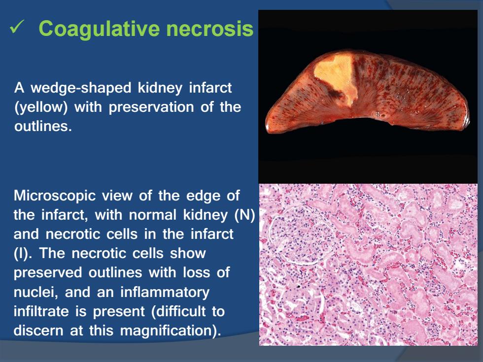
Coagulative necrosis A wedge-shaped kidney infarct (yellow)with preservation of the outlines. Microscopic view of the edge of the infarct,with normal kidney (N) and necrotic cells in the infarct (I).The necrotic cells show preserved outlines with loss of nuclei,and an inflammatory infiltrate is present (difficult to discern at this magnification)
Coagulative necrosis A wedge-shaped kidney infarct (yellow) with preservation of the outlines. Microscopic view of the edge of the infarct, with normal kidney (N) and necrotic cells in the infarct (I). The necrotic cells show preserved outlines with loss of nuclei, and an inflammatory infiltrate is present (difficult to discern at this magnification)

Coagulative necrosis in cardiac infarction
Coagulative necrosis in cardiac infarction

Liquefactive necrosis An infarct in the brain showing dissolution of the tissue. The cystic space contains necrotic cell debris and macrophages filled with phagocytosed material
Liquefactive necrosis An infarct in the brain showing dissolution of the tissue. The cystic space contains necrotic cell debris and macrophages filled with phagocytosed material