Visual Organ EYE For MBBS,Class of 2015
Visual Organ For MBBS, Class of 2015 EYE

Externa Elast Media Sense organs:sense receptor assistant apparatus Sense receptors:sensory units collecting information Exteroceptors:receives stimuli from skin and mucosa Interoceptors:receives stimuli from visceral organs Proprioceptors:receive stimuli from muscles,tendons,joints
Sense organs: sense receptor + assistant apparatus Sense receptors: sensory units collecting information Exteroceptors:receives stimuli from skin and mucosa Interoceptors: receives stimuli from visceral organs Proprioceptors: receive stimuli from muscles, tendons, joints

Visual System Visual Organ Afferent Ragnar Granit Haldan Keffer George Wald Hartline Nerve Sweden USA USA Karolinska Rockefeller Harvard University Institutet University ambridge,MA, Stockholm, New York,NY,USA USA Sweden Visual Cortex David H.Hubel Torsten N.Wiesel USA Sweden
1913 - 1994 1926- 1924- Visual Organ Afferent Nerve Visual Cortex Visual System 1900-1991 1903-1983 1906-1997
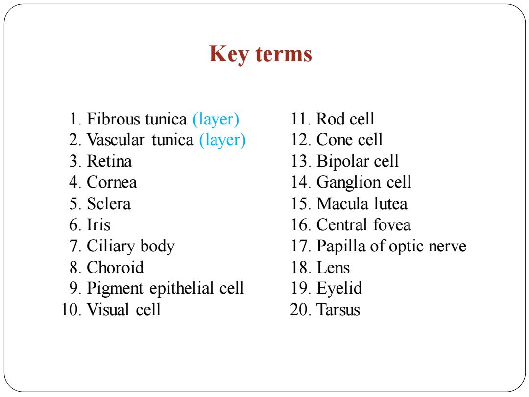
Key terms 1.Fibrous tunica(layer) 11.Rod cell 2.Vascular tunica (layer) 12.Cone cell 3.Retina 13.Bipolar cell 4.Cornea 14.Ganglion cell 5.Sclera 15.Macula lutea 6.Iris 16.Central fovea 7.Ciliary body 17.Papilla of optic nerve 8.Choroid 18.Lens 9.Pigment epithelial cell 19.Eyelid 10.Visual cell 20.Tarsus
Key terms 11. Rod cell 12. Cone cell 13. Bipolar cell 14. Ganglion cell 15. Macula lutea 16. Central fovea 17. Papilla of optic nerve 18. Lens 19. Eyelid 20. Tarsus 1. Fibrous tunica (layer) 2. Vascular tunica (layer) 3. Retina 4. Cornea 5. Sclera 6. Iris 7. Ciliary body 8. Choroid 9. Pigment epithelial cell 10. Visual cell

Eye Basically located inside the orbit(orbital cavity); Consisting of eyeball and accessory organs; Receiving light/image stimuli and transfer them into electrical signals for vision formation; Accessory organs support and protect the eyeballs,and control their movement. 额突 提上睑肌 上直肌 结膜上弯 虹膜 视神经 角膜 -上险 外直肌 下盼 结膜下穹 眼轮匝肌 下直肌下斜肌“烟 右侧眼球及眶腔矢状断
Eye Basically located inside the orbit (orbital cavity); Consisting of eyeball and accessory organs; Receiving light/image stimuli and transfer them into electrical signals for vision formation; Accessory organs support and protect the eyeballs, and control their movement
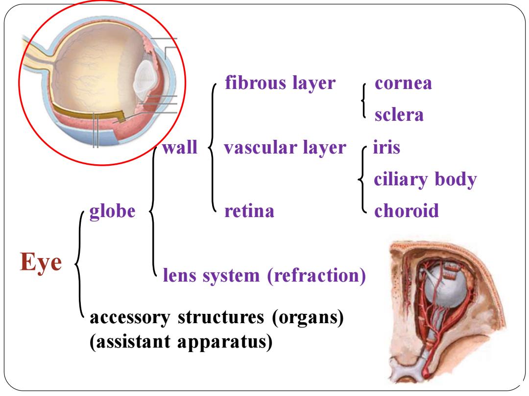
fibrous layer cornea sclera wall vascular layer iris ciliary body globe retina choroid Eye lens system (refraction) accessory structures (organs) (assistant apparatus)
fibrous layer cornea sclera wall vascular layer iris ciliary body globe retina choroid lens system (refraction) accessory structures (organs) (assistant apparatus) Eye
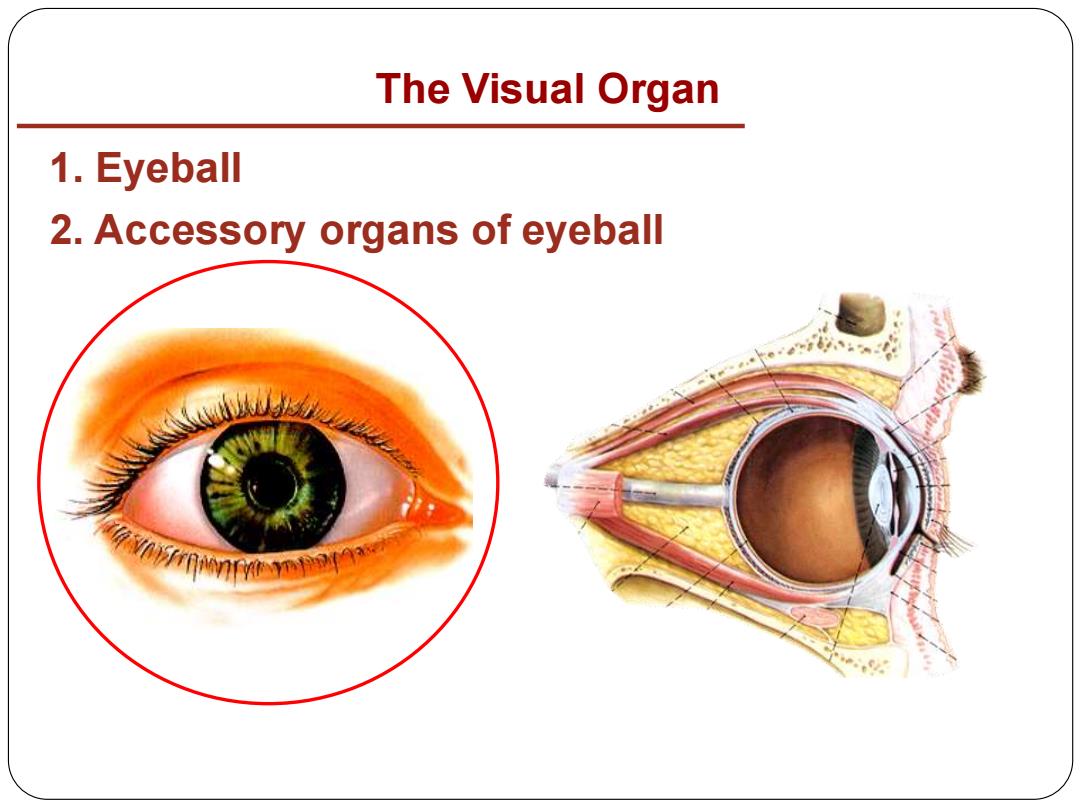
The Visual Organ 1.Eyeball 2.Accessory organs of eyeball 。 VovFrANn91
1. Eyeball 2. Accessory organs of eyeball The Visual Organ
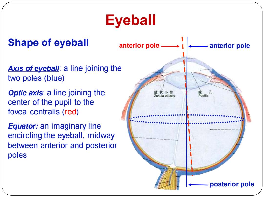
Eyeball Shape of eyeball anterior pole→l anterior pole Axis of eyeball:a line joining the two poles(blue) Optic axis:a line joining the 使状小霜 Zonula ciliaris center of the pupil to the fovea centralis(red) Equator:an imaginary line encircling the eyeball,midway between anterior and posterior poles posterior pole
Eyeball Shape of eyeball anterior pole posterior pole Axis of eyeball: a line joining the two poles (blue) Optic axis: a line joining the center of the pupil to the fovea centralis (red) Equator: an imaginary line encircling the eyeball, midway between anterior and posterior poles anterior pole
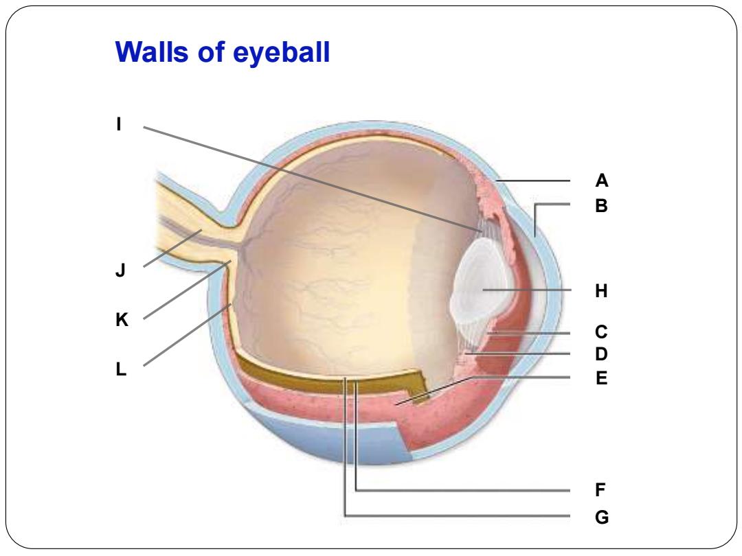
Walls of eyeball m H C a w F G
B C D F E A G H I K J L Walls of eyeball
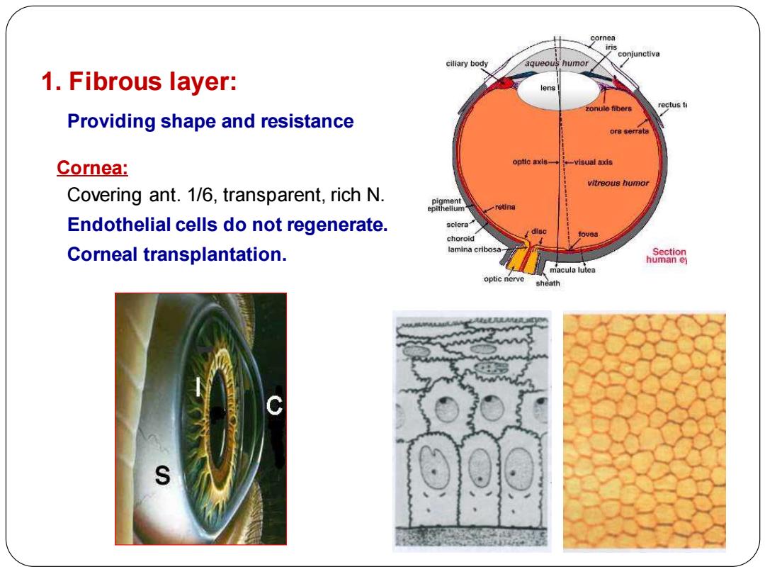
cillary body 1.Fibrous layer: rectus Providing shape and resistance Cornea: visual axis Covering ant.1/6,transparent,rich N. Endothelial cells do not regenerate. sclera choroid Corneal transplantation. lamina cribosa- macula lutea optic nerve sheath
1. Fibrous layer: Providing shape and resistance Cornea: Covering ant. 1/6, transparent, rich N. Endothelial cells do not regenerate. Corneal transplantation