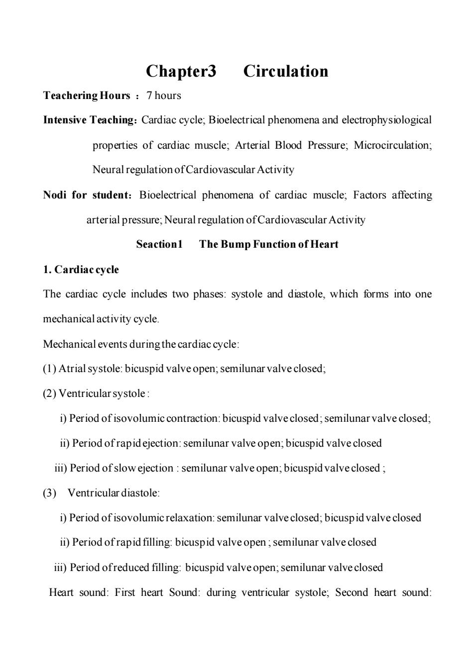
Chapter3 Circulation Teachering Hours 7 hours Intensive Teaching:Cardiac cycle;Bioelectrical phenomena and electrophysiological properties of cardiac muscle;Arterial Blood Pressure;Microcirculation; Neural regulation of Cardiovascular Activity Nodi for student:Bioelectrical phenomena of cardiac muscle;Factors affecting arterial pressure;Neural regulation of Cardiovascular Activity Seaction1 The Bump Function of Heart 1.Cardiac cycle The cardiac cycle includes two phases:systole and diastole,which forms into one mechanical activity cycle. Mechanical events during the cardiac cycle: (1)Atrial systole:bicuspid valve open;semilunar valve closed; (2)Ventricular systole: i)Period of isovolumic contraction:bicuspid valve closed;semilunar valve closed; ii)Period ofrapidejection:semilunar valve open;bicuspid valve closed iii)Period of slow ejection:semilunar valve open;bicuspid valve closed; (3)Ventricular diastole: i)Period of isovolumic relaxation:semilunar valve closed;bicuspid valve closed ii)Period of rapid filling:bicuspid valve open;semilunar valve closed ii)Period ofreduced filling:bicuspid valve open;semilunar valve closed Heart sound:First heart Sound:during ventricular systole;Second heart sound:
Chapter3 Circulation Teachering Hours :7 hours Intensive Teaching:Cardiac cycle; Bioelectrical phenomena and electrophysiological properties of cardiac muscle; Arterial Blood Pressure; Microcirculation; Neural regulation of Cardiovascular Activity Nodi for student:Bioelectrical phenomena of cardiac muscle; Factors affecting arterial pressure; Neural regulation of Cardiovascular Activity Seaction1 The Bump Function of Heart 1. Cardiac cycle The cardiac cycle includes two phases: systole and diastole, which forms into one mechanical activity cycle. Mechanical events during the cardiac cycle: (1) Atrial systole: bicuspid valve open; semilunar valve closed; (2) Ventricular systole : i) Period of isovolumic contraction: bicuspid valve closed;semilunar valve closed; ii) Period of rapid ejection: semilunar valve open; bicuspid valve closed iii) Period of slow ejection : semilunar valve open; bicuspid valve closed ; (3) Ventricular diastole: i) Period of isovolumic relaxation: semilunar valve closed; bicuspid valve closed ii) Period of rapid filling: bicuspid valve open ;semilunar valve closed iii) Period of reduced filling: bicuspid valve open; semilunar valve closed Heart sound: First heart Sound: during ventricular systole; Second heart sound:
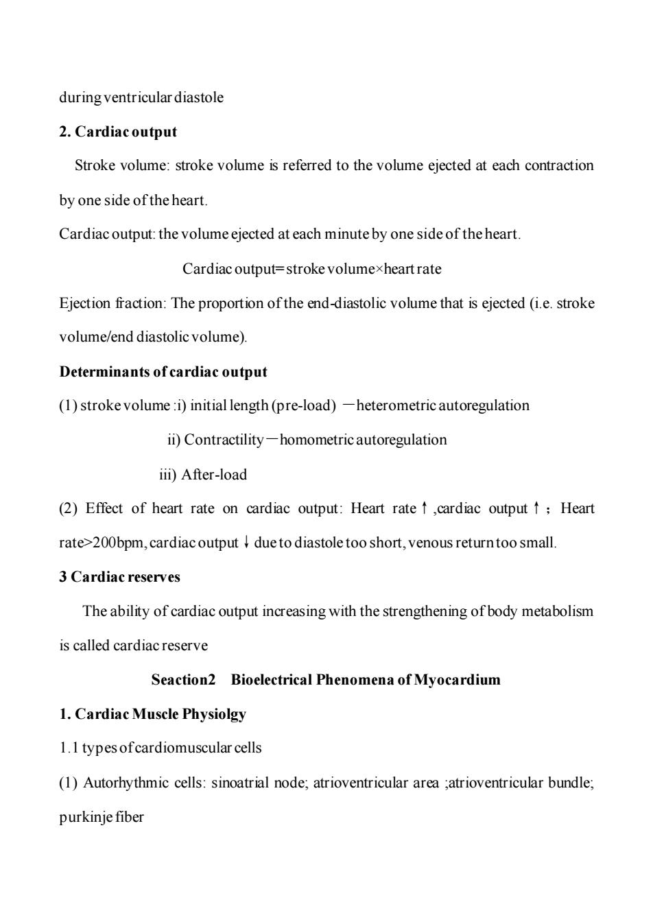
during ventricular diastole 2.Cardiac output Stroke volume:stroke volume is referred to the volume ejected at each contraction by one side of the heart. Cardiac output:the volume ejected at each minute by one side of the heart. Cardiac output=stroke volumexheart rate Ejection fraction:The proportion of the end-diastolic volume that is ejected (i.e.stroke volume/end diastolic volume). Determinants of cardiac output (1)stroke volume:i)initial length(pre-load)-heterometric autoregulation ii)Contractility-homometric autoregulation iii)After-load (2)Effect of heart rate on cardiac output:Heart rate↑,cardiac output↑;Heart rate>200bpm,cardiac output due to diastole too short,venous return too small. 3 Cardiac reserves The ability of cardiac output increasing with the strengthening of body metabolism is called cardiac reserve Seaction2 Bioelectrical Phenomena of Myocardium 1.Cardiac Muscle Physiolgy 1.1 types ofcardiomuscular cells (1)Autorhythmic cells:sinoatrial node;atrioventricular area ;atrioventricular bundle; purkinje fiber
during ventricular diastole 2. Cardiac output Stroke volume: stroke volume is referred to the volume ejected at each contraction by one side of the heart. Cardiac output: the volume ejected at each minute by one side of the heart. Cardiac output= stroke volume×heart rate Ejection fraction: The proportion of the end-diastolic volume that is ejected (i.e. stroke volume/end diastolic volume). Determinants of cardiac output (1) stroke volume :i) initial length (pre-load) -heterometric autoregulation ii) Contractility-homometric autoregulation iii) After-load (2) Effect of heart rate on cardiac output: Heart rate↑,cardiac output↑;Heart rate>200bpm, cardiac output↓due to diastole too short, venous return too small. 3 Cardiac reserves The ability of cardiac output increasing with the strengthening of body metabolism is called cardiac reserve Seaction2 Bioelectrical Phenomena of Myocardium 1. Cardiac Muscle Physiolgy 1.1 types of cardiomuscular cells (1) Autorhythmic cells: sinoatrial node; atrioventricular area ;atrioventricular bundle; purkinje fiber
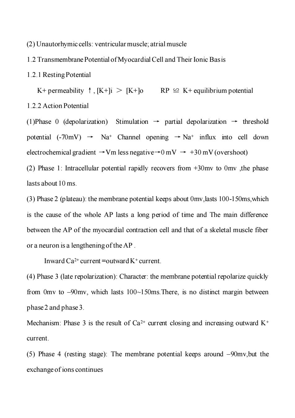
(2)Unautorhymic cells:ventricular muscle;atrial muscle 1.2 Transmembrane Potential of Myocardial Cell and Their Ionic Basis 1.2.1 Resting Potential K+permeability↑,K+]i>K+lo RPK+equilibrium potential 1.2.2 Action Potential (1)Phase 0 (depolarization)Stimulation partial depolarization threshold potential (-70mV)Na+Channel opening -Na+influx into cell down electrochemical gradient→Vm less negative-→0mV→+30mV(overshoot) (2)Phase 1:Intracellular potential rapidly recovers from +30mv to Omv ,the phase lasts about 10 ms. (3)Phase 2(plateau):the membrane potential keeps about 0mv,lasts 100-150ms,which is the cause of the whole AP lasts a long period of time and The main difference between the AP of the myocardial contraction cell and that of a skeletal muscle fiber or a neuron is a lengthening of the AP. Inward Ca2+current =outward K+current. (4)Phase 3(late repolarization):Character:the membrane potential repolarize quickly from Omv to -90mv,which lasts 100~150ms.There,is no distinct margin between phase 2 and phase 3 Mechanism:Phase 3 is the result of Ca2+current closing and increasing outward K+ current. (5)Phase 4 (resting stage):The membrane potential keeps around -90mv,but the exchange ofions continues
(2) Unautorhymic cells: ventricular muscle; atrial muscle 1.2 Transmembrane Potential of Myocardial Cell and Their Ionic Basis 1.2.1 Resting Potential K+ permeability ↑, [K+]i > [K+]o RP ≌ K+ equilibrium potential 1.2.2 Action Potential (1)Phase 0 (depolarization) Stimulation → partial depolarization → threshold potential (-70mV) → Na+ Channel opening → Na+ influx into cell down electrochemical gradient →Vm less negative→0 mV → +30 mV (overshoot) (2) Phase 1: Intracellular potential rapidly recovers from +30mv to 0mv ,the phase lasts about 10 ms. (3) Phase 2 (plateau): the membrane potential keeps about 0mv,lasts 100-150ms,which is the cause of the whole AP lasts a long period of time and The main difference between the AP of the myocardial contraction cell and that of a skeletal muscle fiber or a neuron is a lengthening of the AP . Inward Ca2+ current =outward K+ current. (4) Phase 3 (late repolarization): Character: the membrane potential repolarize quickly from 0mv to –90mv, which lasts 100~150ms.There, is no distinct margin between phase 2 and phase 3. Mechanism: Phase 3 is the result of Ca2+ current closing and increasing outward K+ current. (5) Phase 4 (resting stage): The membrane potential keeps around –90mv,but the exchange of ions continues
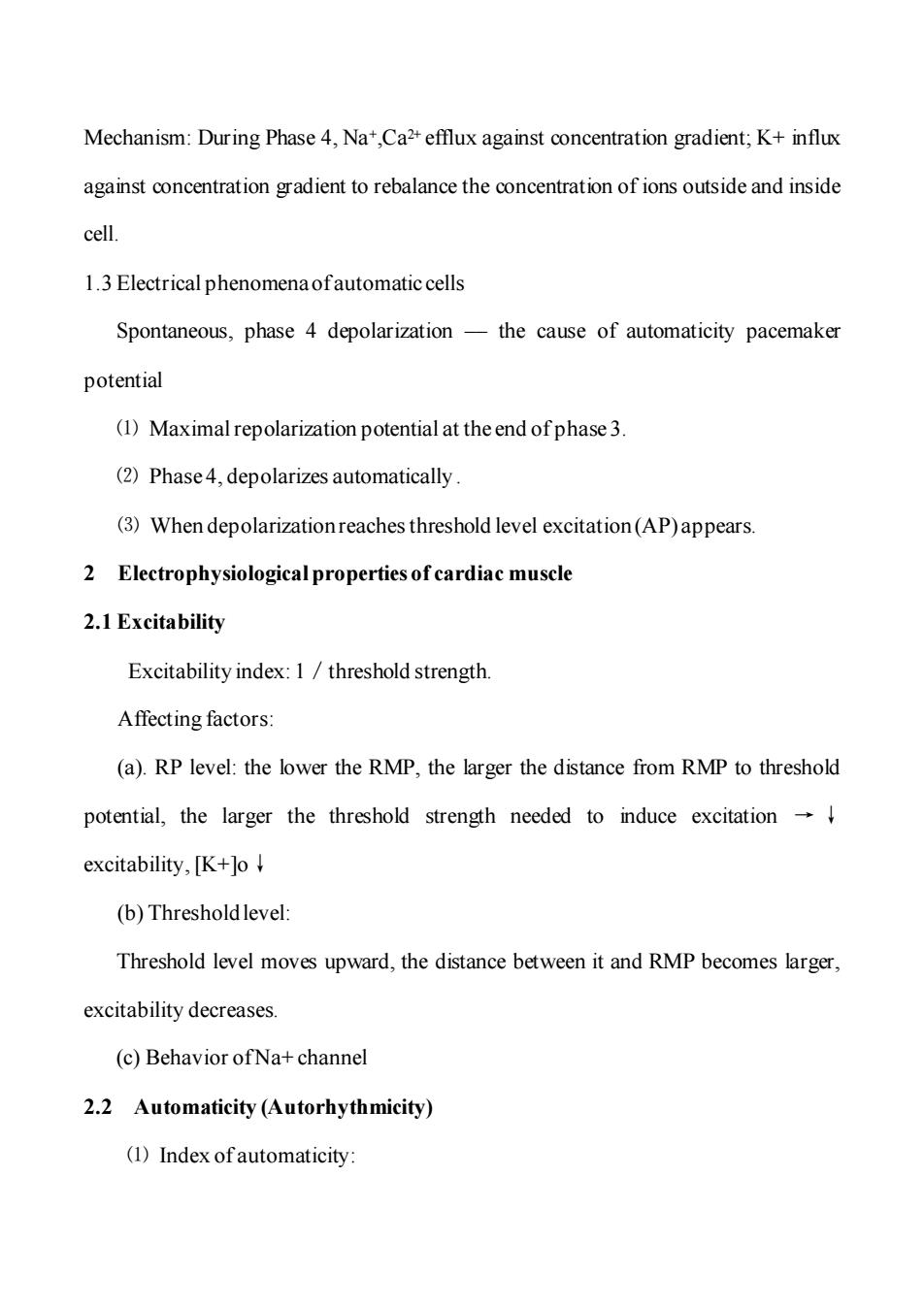
Mechanism:During Phase 4,Na,Ca2+efflux against concentration gradient;K+influx against concentration gradient to rebalance the concentration of ions outside and inside cell. 1.3 Electrical phenomena ofautomatic cells Spontaneous,phase 4 depolarization-the cause of automaticity pacemaker potential (1)Maximal repolarization potential at the end of phase 3. (2)Phase4,depolarizes automatically. (3)When depolarizationreaches threshold level excitation(AP)appears 2 Electrophysiological properties of cardiac muscle 2.1 Excitability Excitability index:1 threshold strength. Affecting factors: (a).RP level:the lower the RMP,the larger the distance from RMP to threshold potential,the larger the threshold strength needed to induce excitation excitability,[K+lo (b)Thresholdlevel: Threshold level moves upward,the distance between it and RMP becomes larger, excitability decreases. (c)Behavior ofNa+channel 2.2 Automaticity (Autorhythmicity) (1)Index of automaticity:
Mechanism: During Phase 4, Na+,Ca2+ efflux against concentration gradient; K+ influx against concentration gradient to rebalance the concentration of ions outside and inside cell. 1.3 Electrical phenomena of automatic cells Spontaneous, phase 4 depolarization –– the cause of automaticity pacemaker potential ⑴ Maximal repolarization potential at the end of phase 3. ⑵ Phase 4, depolarizes automatically . ⑶ When depolarization reaches threshold level excitation (AP) appears. 2 Electrophysiological properties of cardiac muscle 2.1 Excitability Excitability index: 1/threshold strength. Affecting factors: (a). RP level: the lower the RMP, the larger the distance from RMP to threshold potential, the larger the threshold strength needed to induce excitation → ↓ excitability, [K+]o↓ (b) Threshold level: Threshold level moves upward, the distance between it and RMP becomes larger, excitability decreases. (c) Behavior of Na+ channel 2.2 Automaticity (Autorhythmicity) ⑴ Index of automaticity:
frequency of discharge of pacemaker cell in S-A node.100/min:dominant pacemaker A-V junction 50/min, Purkinje fiber 25/min Latent or subordinary pacemaker (2)Factors determining automaticity Frequency of excitation of pacemaker cell determinates the time for maximum diastolic potential to reach threshold potential. a.Rate ofspontaneous,phase 4 depolarization.B receptor activation,If t HR t b.Maximum diastolic potential level,gK+HR c.Threshold potential level 2.3 Conductivity 2.3.1.Index ofconductivity-speed of conduction of AP 2.3.2.Factors determining conductivity ofcardiac muscle a.Speed and amplitude ofphase 0 depolarization (a)↑speed of phase 0 depolarization→↑rate of generation of local current→I time for depolarization to reach threshold potential-conductivity t (b)↑amplitude of phase 0 depolarization→↑amplitude of local current→t distance ofdepolarization ofnearby membrane-t conductivity (c)Speed and amplitude ofphase0 depolarization is determined by Vm More negative RP→↑speed of Na+channel opening→↑speed of phase depolarizationt speed of local current stimulation to reach to threshold potential
frequency of discharge of pacemaker cell in S-A node. 100/min: dominant pacemaker A-V junction 50/min, Purkinje fiber 25/min Latent or subordinary pacemaker ⑵ Factors determining automaticity Frequency of excitation of pacemaker cell determinates the time for maximum diastolic potential to reach threshold potential. a. Rate of spontaneous, phase 4 depolarization.βreceptor activation, If↑ HR↑ b. Maximum diastolic potential level , gK+↑ HR↓ c. Threshold potential level 2.3 Conductivity 2.3.1. Index of conductivity – speed of conduction of AP 2.3.2. Factors determining conductivity of cardiac muscle a. Speed and amplitude of phase 0 depolarization (a) ↑ speed of phase 0 depolarization → ↑rate of generation of local current →↓ time for depolarization to reach threshold potential → conductivity ↑ (b)↑amplitude of phase 0 depolarization → ↑amplitude of local current → ↑ distance of depolarization of nearby membrane → ↑conductivity. (c) Speed and amplitude of phase 0 depolarization is determined by Vm More negative RP → ↑speed of Na+ channel opening → ↑speed of phase depolarization → ↑speed of local current stimulation to reach to threshold potential

→↑speed of conduction b.excitability ofnearby membrane area-conductivity Local current stimulus conducts to area which is in effective refractory period of premature potential.The stimulus can't induce a new AP and conduction block occurs. 2.4 Contractility 3 Electrocardiogram (ECG) The synchronized depolarizations spreading through the heart cause currents that establish field potential,whose differences can be amplified and detected by electrodes placed on the body surface.The record produced is called electrocardiogram. A normal ECG is composed of P wave,QRS complex,and T wave.The P wave represents the depolarization process of both right and left atriums.The QRS complex is caused by the depolarization of both ventricles and the T wave is generated by the repolarization process of the ventricles.The P-R interval is a measure of the time from the onset of atrial excitation to the onset of ventricular activation.The prolongation of P-R interval always indicates the disturbance in A-V conduction. Seaction3 Vascular Physiolgy 1 Functional properties of different blood vessels 1)Artery:Aortaand large artery;Pressure reservoir vessels 2)Capillary vessels:exchange vessels;Exchange of substances between blood and interstitial fluid 3)Venous Vessels,capacitance vessels:large vein,vena cava
→ ↑speed of conduction b. ↓excitability of nearby membrane area → ↓conductivity Local current stimulus conducts to area which is in effective refractory period of premature potential. The stimulus can’t induce a new AP and conduction block occurs. 2.4 Contractility 3 Electrocardiogram (ECG) The synchronized depolarizations spreading through the heart cause currents that establish field potential, whose differences can be amplified and detected by electrodes placed on the body surface. The record produced is called electrocardiogram. A normal ECG is composed of P wave, QRS complex, and T wave. The P wave represents the depolarization process of both right and left atriums. The QRS complex is caused by the depolarization of both ventricles and the T wave is generated by the repolarization process of the ventricles. The P-R interval is a measure of the time from the onset of atrial excitation to the onset of ventricular activation. The prolongation of P-R interval always indicates the disturbance in A-V conduction. Seaction3 VascularPhysiolgy 1 Functional properties of different blood vessels 1)Artery: Aorta and large artery; Pressure reservoir vessels 2) Capillary vessels: exchange vessels; Exchange of substances between blood and interstitial fluid 3)Venous Vessels, capacitance vessels: large vein, vena cava

2 Blood Pressure:the force exerted by the blood against any unit area of the vessel wall 2.1 Arterial Blood Pressure Systolic Pressure Diastolic Pressure Pulse Pressure MAP=DP+1/3(pulseP) 2.2 Factors affecting arterial pressure (1)stoke volume (2)heartrate (3)peripheral resistance (4)aorta large artery (5)circulatory blood flow 2.3 Venous pressure and venous return 2.3.1 Central venous pressure Affect factors:A.Heart pump action B.Venous return velocity 2.3.2 Venous returnand affecting factors A.Mean circulatory filling pressure B.Cardiac contractility C.Sympathetic nerve 3 Microcirculation,Interstitial fluid and Lymph 3.1 Microcirculation Microcirculation is the blood circulation between arteriole and veinule.The
2 Blood Pressure: the force exerted by the blood against any unit area of the vessel wall 2.1 Arterial Blood Pressure Systolic Pressure Diastolic Pressure Pulse Pressure MAP = DP + 1/3 (pulse P) 2.2 Factors affecting arterial pressure (1) stoke volume (2) heart rate (3) peripheralresistance (4) aorta large artery (5)circulatory blood flow 2.3 Venous pressure and venous return 2.3.1 Central venous pressure Affect factors:A. Heart pump action B. Venous return velocity 2.3.2 Venous return and affecting factors A. Mean circulatory filling pressure B. Cardiac contractility C. Sympathetic nerve 3 Microcirculation,Interstitial fluid and Lymph 3.1 Microcirculation Microcirculation is the blood circulation between arteriole and veinule.The
microcirculation is the place where the blood and interstitial fluid to carry out the exchange of substances. (1)Thoroughfare or preferential channel. (2)A-V anastomosis or A-v shunt. (3)Arteriole→metareriole→precapillary sphincter→true capillary→ venule. 3.2 Formation of interstitial fluid The filtration and reabsorption of fluid through the capillary wall is determined by the difference of the strength offiltrationand reabsorption. EP=(Pc+if)-(p+Pif) Factors affecting the formation ofinterstitial fluid: (1)Capillary hydrostatic pressure. (2)Colloid osmotic pressure. (3)Lymph return. (4)Capillary permeability. 3.3 Formation and return oflymph physiological function:i)Reclaiming protein and absorbing nourishment ii)Cleaning the RBC.bacterium and foreign substance iii)Balancing the formation and absorption ofinterstitial fluid Seaction4 Regulation of Cardiovascular Activity 1 Neural regulation of Cardiovascular Activity 1.1 Cardiac innervation
microcirculation is the place where the blood and interstitial fluid to carry out the exchange of substances. (1) Thoroughfare or preferential channel. (2) A-V anastomosis or A-v shunt. (3) Arteriole → metareriole → precapillary sphincter → true capillary → venule. 3.2 Formation of interstitial fluid The filtration and reabsorption of fluid through the capillary wall is determined by the difference of the strength of filtration and reabsorption. EP= ( Pc + if ) – ( p + Pif ) Factors affecting the formation of interstitial fluid: (1) Capillary hydrostatic pressure. (2) Colloid osmotic pressure. (3) Lymph return. (4) Capillary permeability. 3.3 Formation and return of lymph physiological function: i)Reclaiming protein and absorbing nourishment ii)Cleaning the RBC、bacterium and foreign substance iii) Balancing the formation and absorption of interstitial fluid Seaction4 Regulation of Cardiovascular Activity 1 Neural regulation of Cardiovascular Activity 1.1 Cardiac innervation

(1)Effects ofvagal nerve Vagal nerve ending→ACh.→binds to M cholinergic receptor→t permeability to K+results in:automaticity ofS-A node ↓contractility due to:↑K+efflux at phase3 repolarization→↓AP duration→ Ca2+influx↓→[Ca2+]i↓;ACh inhibits Caz2+influx→[Ca2+]i↓→↓ contractility and conductivity (2)Effects of cardiac sympathetic nerve: Cardiac sympathetic nerve ending-noradrenaline -binds to B-adrenergic receptor→↑permeability to Ca2+leads to: ↑Automaticity ↑Conductivity ↑Contractility 1.2 Innervation ofblood vessels (1)Vasoconstriction fiber (2)Vasodilationnerve fibebs 1)Sympathetic vasodilation never fibers 2)Parasymp.vasodilation never fibers.(ACh) 3)Non-cholinergic,non-adrenergic fibers 1.3 Cardiovascular center 1.3.1 Cardiovascular center in medulla oblongata (1)Rostral ventrolateral medulla(RVLM) (2)Caudal ventrolateral medulla
(1) Effects of vagal nerve Vagal nerve ending → ACh. → binds to M cholinergic receptor → ↑ permeability to K+ results in:↓automaticity of S-A node ↓contractility due to : ↑K+ efflux at phase 3 repolarization →↓AP duration → Ca2+ influx ↓→ [Ca2+]i↓ ; ACh inhibits Ca2+ influx → [Ca2+]i ↓→ ↓ contractility and ↓conductivity (2) Effects of cardiac sympathetic nerve: Cardiac sympathetic nerve ending → noradrenaline → binds to β-adrenergic receptor→↑permeability to Ca2+ leads to: ↑Automaticity ↑Conductivity ↑Contractility 1.2 Innervation of blood vessels (1) Vasoconstriction fiber (2) Vasodilation nerve fibebs 1) Sympathetic vasodilation never fibers 2) Parasymp. vasodilation never fibers. (ACh) 3)Non-cholinergic, non-adrenergic fibers 1.3 Cardiovascular center 1.3.1 Cardiovascular center in medulla oblongata (1) Rostral ventrolateral medulla(RVLM) (2) Caudal ventrolateral medulla
(3)Nucleus ofsolitary tract (4)Cardiac vagal center 1.3.2 Anterior hypothalamus (AH) 1.3.3 Cerebral cortex 1.4 Cardiovascular regulatory mechanism 1.4.1 Arterial baroreflexes: (1).Baroreceptor is located in adventitia of carotid sinus and aortic arch (2)Afferent nerve:Buffer nerves When artery pressure rises,baroreceptor reflex will be aroused,the heart rate and peripheral resistance decrease,then the blood pressure falls back to normal.This reflex is also called depressor reflex. Functional significances of arterial baroreflex:i)To keep arterial pressure at normal level;ii)To stabilize arterial pressure and prevent arterial pressure from large fluctuation 1.4.2 Chemoreceptor reflex (1)Respiratory depthand frequency increases; (2)Vasoconstriction in skeletal M,splanchnic viscera,kidney. (3)Blood flow in brain and liver increases.BP inceases (4)↑HR,↑cardiac output due to a.respirationrate and depth b.t catecholamine from adrenal medulla 2 Humoral regulation
(3) Nucleus of solitary tract (4) Cardiac vagal center 1.3.2 Anterior hypothalamus(AH) 1.3.3 Cerebral cortex 1.4 Cardiovascular regulatory mechanism 1.4.1 Arterial baroreflexes: (1). Baroreceptor is located in adventitia of carotid sinus and aortic arch (2) Afferent nerve: Buffer nerves When artery pressure rises, baroreceptor reflex will be aroused, the heart rate and peripheral resistance decrease, then the blood pressure falls back to normal. This reflex is also called depressor reflex. Functional significances of arterial baroreflex: i)To keep arterial pressure at normal level; ii)To stabilize arterial pressure and prevent arterial pressure from large fluctuation 1.4.2 Chemoreceptor reflex (1) Respiratory depth and frequency increases; (2) Vasoconstriction in skeletal M, splanchnic viscera, kidney. (3) Blood flow in brain and liver increases. BP inceases (4)↑HR, ↑cardiac output due to a.↑respiration rate and depth b.↑catecholamine from adrenal medulla. 2 Humoral regulation