Chapter 1 Celluar Physiology Teachering Hours 8 hours Intensive Teaching:structure of cell membrane and transmembrane transport; Transmembrane signal transduction;electrical phenomena of the cells;contraction function ofthe skeleton muscle Nodi for student:The mechanism of resting potential and action potential;The contraction mechanism ofskeleton muscle Seaction1 Structure of Cell Membrane and Membrane Transport 1.Composition and structure of cell membrane The approximate composition of the cell membrane proteins,55%;phospholipids,25%;cholesterol,13%: other lipids,4;and carbohydrates.3%.The fluid mosaic model is used to elucidate the structure of membrane,which was first expounded by Singer and Nicholson in 1972. 1.1 fluid mosaic model The cell membraneconsists of a double layer of lipid meolecules in which globular proteins are embedded. 1.1.1 Lipid bilayer The basic lipid bilayer is composed of phospholipids molecules which are amphipathic molecules.One end of each phospholipids has a charged region and is hydrophilic.The other end consists of twolong fatty acid chains and is hydrophobic.The phospholipids in cell membrane are organized into a bi-molecular layer with the hydrophobic fatty acid chains in the middle.The hydrophilic regions of
Chapter 1 Celluar Physiology Teachering Hours :8 hours Intensive Teaching:structure of cell membrane and transmembrane transport; Transmembrane signal transduction; electrical phenomena of the cells; contraction function of the skeleton muscle Nodi for student:The mechanism of resting potential and action potential; The contraction mechanism of skeleton muscle Seaction1 Structure of Cell Membrane and Membrane Transport 1. Composition and structure of cell membrane The approximate composition of the cell membrane is proteins,55%;phospholipids,25%;cholesterol,13%; other lipids,4;and carbohydrates,3%.The fluid mosaic model is used to elucidate the structure of membrane,which was first expounded by Singer and Nicholson in 1972. 1.1 fluid mosaic model The cell membraneconsists of a double layer of lipid meolecules in which globular proteins are embedded. 1.1.1 Lipid bilayer The basic lipid bilayer is composed of phospholipids molecules which are amphipathic molecules.One end of each phospholipids has a charged region and is hydrophilic.The other end consists of twolong fatty acid chains and is hydrophobic .The phospholipids in cell membrane are organized into a bi-molecular layer with the hydrophobic fatty acid chains in the middle.The hydrophilic regions of
the phospholipids are oriented toward the surfaces of the membrane as a result of their attraction to the polar water molecules in the extracellular and cytosol. 1.1.2 Membrane proteins:(1)integral proteins (2)peripheral proteins 1.1.3 carbohydrates:Play important roles in enabling cells to identify and interact with each other. 2.Transmembrane transport 2.1 passivetransport The movement of substances across the membrane by down their concentration or electrochemical gradient without consumption of metabolic energy. (1)simple diffusion: The movement of small lipid soluble substances directly through the cell membrane down the concentration gradient. (2)Facilitated diffusion The diffusion of small water-soluble substances through the cell membrane with the aid of particular membrane transport proteins.These transport proteins are classified as channel proteins or carrier proteins. 1characteristics ofcarrier-mediated facilitated diffusion: a.specificity b.saturation competition 2characteristics of channel-mediated facilitated diffusion: 1)Voltate-gated channel: 2)Chemically-gated channel: 2.2 activetransport
the phospholipids are oriented toward the surfaces of the membrane as a result of their attraction to the polar water molecules in the extracellular and cytosol. 1.1.2 Membrane proteins:(1)integral proteins (2)peripheral proteins 1.1.3 carbohydrates: Play important roles in enabling cells to identify and interact with each other. 2. Transmembrane transport 2.1 passive transport The movement of substances across the membrane by down their concentration or electrochemical gradient without consumption of metabolic energy. (1) simple diffusion: The movement of small lipid soluble substances directly through the cell membrane down the concentration gradient. (2) Facilitated diffusion The diffusion of small water-soluble substances through the cell membrane with the aid of particular membrane transport proteins.These transport proteins are classified as channel proteins or carrier proteins. ①characteristics of carrier-mediated facilitated diffusion: a. specificity b. saturation c. competition ②characteristics of channel-mediated facilitated diffusion: 1)Voltate-gated channel: 2)Chemically-gated channel: 2.2 active transport
The movement of substances across the membrane occurs against the concentration gradient or electrochemical gradient with the necessity of consumption of metabolic energy characteristics (1 movement against the concentration gradient or electrochemical gradient (2)consumption ofenergy 2.2.1 Primary activetransport Making direct use of enery derived from ATP to transport the ions across to the cell membrane. 2.2.2:Secondary active transport The ion gradients established by primary active transport permits the transport of other substances against their concentration gradients. 2.2.3 Sodium--potassium pump (1Making the Na+and K+gradients across the cell membrane. (2)Providing energy for secondary activetransport 2.3 exocytosis and endocytosis (1)Endocytosis:Very large particles enter the cell by a specialized function of the cell membrane called endocytosis.The principle forms of endocytosis are pinocytosis and phagocytosis. (2)Exocytosis:A stimulus to secrete causes the intracellular vesicles to fuse with the plasma membrane and to release the vesicles contents is called exocytosis. 3 Signal transduction
The movement of substances across the membrane occurs against the concentration gradient or electrochemical gradient with the necessity of consumption of metabolic energy. characteristics : (1 ) movement against the concentration gradient or electrochemical gradient (2)consumption of energy 2.2.1 Primary active transport Making direct use of enery derived from ATP to transport the ions across to the cell membrane. 2.2.2:Secondary active transport The ion gradients established by primary active transport permits the transport of other substances against their concentration gradients. 2.2.3 Sodium--potassium pump (1)Making the Na+ and K+ gradients across the cell membrane. (2)Providing energy for secondary active transport 2.3 exocytosis and endocytosis (1) Endocytosis: Very large particles enter the cell by a specialized function of the cell membrane called endocytosis. The principle forms of endocytosis are pinocytosis and phagocytosis. (2) Exocytosis: A stimulus to secrete causes the intracellular vesicles to fuse with the plasma membrane and to release the vesicles contents is called exocytosis. 3 Signal transduction
Signal transduction refers to the processes by which intercellular messengers (such as neurotransmitters,hormones and cytokines)which bind to specific receptors on or in the target cell,are converted into biochemical and/or electrical signals within that cell.In turn,such signals can modify cellular function in different ways.Three general patterns of signal transduction occur in almost all mammalian cells.One is signal transduction mediated by G-protein coupled receptor.G-protein serve as intermediaries between receptor and the enzymes or ion channels that activated by agonist binding.The second is signal transduction mediated by ion channel-coupled receptors which help regulate the intracellular concentration of specific ions.The third pattern,some membrane receptors are protein kinases called tyrosine protein kinase that are activated directly by agonist binding. Several basic concepts for the signal transduction first messenger:including the lipid-insoluble messenger and lipid-soluble messenger. Second messenger:it is the signal molecule that produced in interior of cell and can transfer the informations carried by first messenger to regulate the protein kinases and ion channels,and to initiate the physiological and biochemical reactions.For example: cAMP,cGMP,IP3,DG,Ca2+etc. protein kinase:It can phosphorylate specific proteins,triggering biochemical reaction that lead to the cell's response to the hormone Signal transduction mediated by G-protein-linked receptor pathway Activation of the receptor by the binding ofthe first messenter allows the receptor
Signal transduction refers to the processes by which intercellular messengers (such as neurotransmitters, hormones and cytokines) which bind to specific receptors on or in the target cell, are converted into biochemical and/or electrical signals within that cell. In turn, such signals can modify cellular function in different ways. Three general patterns of signal transduction occur in almost all mammalian cells. One is signal transduction mediated by G-protein coupled receptor. G-protein serve as intermediaries between receptor and the enzymes or ion channels that activated by agonist binding. The second is signal transduction mediated by ion channel-coupled receptors which help regulate the intracellular concentration of specific ions. The third pattern, some membrane receptors are protein kinases called tyrosine protein kinase that are activated directly by agonist binding. Several basic concepts for the signal transduction first messenger: including the lipid-insoluble messenger and lipid-soluble messenger. Second messenger: it is the signal molecule that produced in interior of cell and can transfer the informations carried by first messenger to regulate the protein kinases and ion channels, and to initiate the physiological and biochemical reactions. For example: cAMP, cGMP, IP3, DG, Ca2+ etc. protein kinase: It can phosphorylate specific proteins, triggering biochemical reaction that lead to the cell’s response to the hormone Signal transduction mediated by G-protein-linked receptor pathway Activation of the receptor by the binding of the first messenter allows the receptor
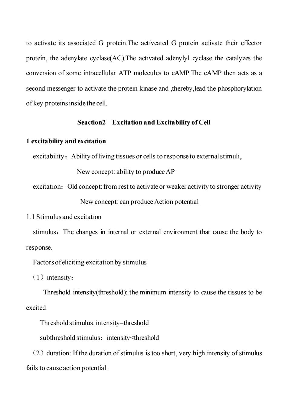
to activate its associated G protein.The activeated G protein activate their effector protein,the adenylate cyclase(AC).The activated adenylyl cyclase the catalyzes the conversion of some intracellular ATP molecules to cAMP.The cAMP then acts as a second messenger to activate the protein kinase and ,thereby,lead the phosphorylation ofkey proteins inside the cell. Seaction2 Excitation and Excitability of Cell 1 excitability and excitation excitability:Ability ofliving tissues or cells to response to external stimuli, New concept:ability to produce AP excitation:Old concept:from rest to activate or weaker activity to stronger activity New concept:can produce Action potential 1.1 Stimulus and excitation stimulus:The changes in internal or external environment that cause the body to response. Factors ofeliciting excitation by stimulus (1)intensity: Threshold intensity(threshold):the minimum intensity to cause the tissues to be excited. Threshold stimulus:intensity=threshold subthreshold stimulus:intensity<threshold (2)duration:If the duration of stimulus is too short,very high intensity of stimulus fails to cause action potential
to activate its associated G protein.The activeated G protein activate their effector protein, the adenylate cyclase(AC).The activated adenylyl cyclase the catalyzes the conversion of some intracellular ATP molecules to cAMP.The cAMP then acts as a second messenger to activate the protein kinase and ,thereby,lead the phosphorylation of key proteins inside the cell. Seaction2 Excitation and Excitability of Cell 1 excitability and excitation excitability:Ability of living tissues or cells to response to external stimuli, New concept: ability to produce AP excitation:Old concept: from rest to activate or weaker activity to stronger activity New concept: can produce Action potential 1.1 Stimulus and excitation stimulus:The changes in internal or external environment that cause the body to response. Factors of eliciting excitation by stimulus (1)intensity: Threshold intensity(threshold): the minimum intensity to cause the tissues to be excited. Threshold stimulus: intensity=threshold subthreshold stimulus:intensity<threshold (2)duration: If the duration of stimulus is too short, very high intensity of stimulus fails to cause action potential
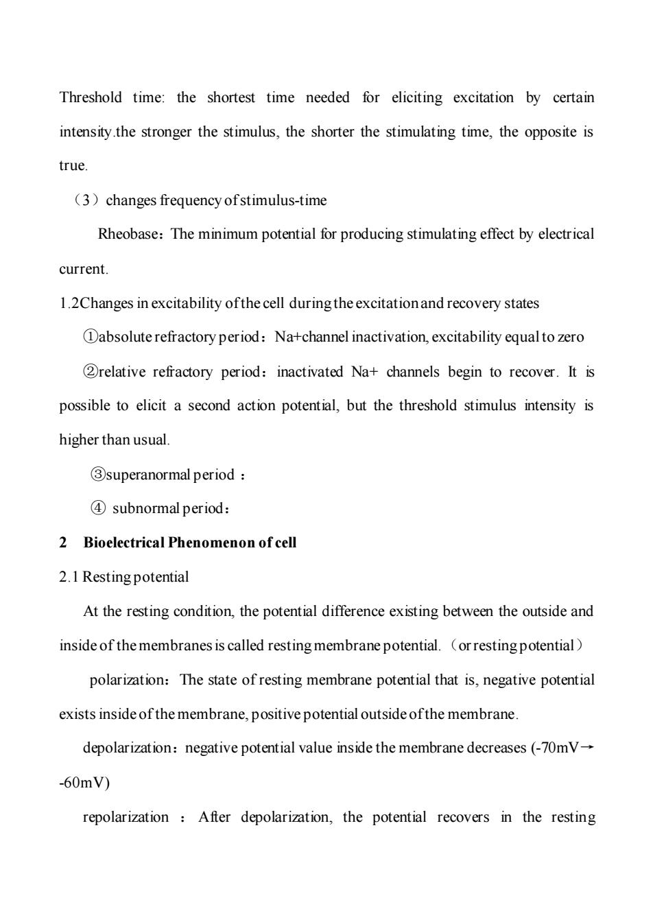
Threshold time:the shortest time needed for eliciting excitation by certain intensity.the stronger the stimulus,the shorter the stimulating time,the opposite is true. (3)changes frequency ofstimulus-time Rheobase:The minimum potential for producing stimulating effect by electrical current. 1.2Changes in excitability ofthe cell during the excitation and recovery states Dabsolute refractory period:Na+channel inactivation,excitability equalto zero 2relative refractory period:inactivated Na+channels begin to recover.It is possible to elicit a second action potential,but the threshold stimulus intensity is higher than usual. 3superanormal period ④subnormal period: 2 Bioelectrical Phenomenon of cell 2.1 Resting potential At the resting condition,the potential difference existing between the outside and inside of the membranes is called resting membrane potential.(or resting potential) polarization:The state of resting membrane potential that is,negative potential exists inside of the membrane,positive potential outside ofthe membrane. depolarization:negative potential value inside the membrane decreases (-70mV- -60mV) repolarization After depolarization,the potential recovers in the resting
Threshold time: the shortest time needed for eliciting excitation by certain intensity.the stronger the stimulus, the shorter the stimulating time, the opposite is true. (3)changes frequency of stimulus-time Rheobase:The minimum potential for producing stimulating effect by electrical current. 1.2Changes in excitability of the cell during the excitation and recovery states ①absolute refractory period:Na+channel inactivation, excitability equal to zero ②relative refractory period:inactivated Na+ channels begin to recover. It is possible to elicit a second action potential, but the threshold stimulus intensity is higher than usual. ③superanormal period : ④ subnormal period: 2 Bioelectrical Phenomenon of cell 2.1 Resting potential At the resting condition, the potential difference existing between the outside and inside of the membranes is called resting membrane potential.(or resting potential) polarization:The state of resting membrane potential that is, negative potential exists inside of the membrane, positive potential outside of the membrane. depolarization:negative potential value inside the membrane decreases (-70mV→ -60mV) repolarization : After depolarization, the potential recovers in the resting

membrane potential direction. hyperarization negative potential value inside the membrane increases(-70mV →-90mV) 2.2 Action potential When the cell or the nerve fiber is stimulated by certain intensity of electrical stimuli,the membrane potential happens a sudden change from the normal resting negative membrane potential to a positive potential and then ends with an almost equally rapid change back to the negative potential,that is the action potential Action potential include three stage: (1)depolarization:The normal polarized"state of-70mV rising rapidly in positive direction. (2)contrapolarization:The membrane potential beyond the zero level and becomes somewhat positive (3)repolarization:The membrane potential returns back to the normal negative potential Characteristics of AP (1)all or none rule:All action potentials in a given cell are the same size regardless of their amplitude of stimulus and this phenomenon is called all-or-non rule (2)AP can transfer to surrounding site 2.3 Mechanism ofGenerating Bioelectricity 2.3.1 mechanism ofresting potential
membrane potential direction. hyperarization :negative potential value inside the membrane increases( -70mV → -90mV) 2.2 Action potential When the cell or the nerve fiber is stimulated by certain intensity of electrical stimuli, the membrane potential happens a sudden change from the normal resting negative membrane potential to a positive potential and then ends with an almost equally rapid change back to the negative potential, that is the action potential Action potential include three stage: (1)depolarization: The normal “polarized” state of -70mV rising rapidly in positive direction. (2)contrapolarization: The membrane potential beyond the zero level and becomes somewhat positive (3)repolarization: The membrane potential returns back to the normal negative potential Characteristics of AP (1)all or none rule: All action potentials in a given cell are the same size regardless of their amplitude of stimulus and this phenomenon is called all-or-non rule. (2) AP can transfer to surrounding site 2.3 Mechanism of Generating Bioelectricity 2.3.1 mechanism of resting potential
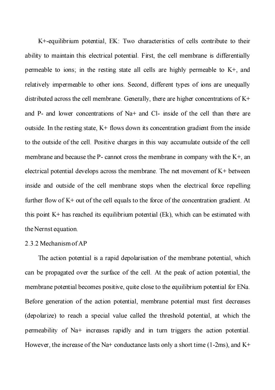
K+-equilibrium potential,EK:Two characteristics of cells contribute to their ability to maintain this electrical potential.First,the cell membrane is differentially permeable to ions;in the resting state all cells are highly permeable to K+,and relatively impermeable to other ions.Second,different types of ions are unequally distributed across the cell membrane.Generally,there are higher concentrations of K+ and P-and lower concentrations of Na+and Cl-inside of the cell than there are outside.In the resting state,K+flows down its concentration gradient from the inside to the outside of the cell.Positive charges in this way accumulate outside of the cell membrane and because the P-cannot cross the membrane in company with the K+,an electrical potential develops across the membrane.The net movement of K+between inside and outside of the cell membrane stops when the electrical force repelling further flow of K+out of the cell equals to the force of the concentration gradient.At this point K+has reached its equilibrium potential(Ek),which can be estimated with the Nernst equation. 2.3.2 Mechanism ofAP The action potential is a rapid depolarisation of the membrane potential,which can be propagated over the surface of the cell.At the peak of action potential,the membrane potential becomes positive,quite close to the equilibrium potential for ENa. Before generation of the action potential,membrane potential must first decreases (depolarize)to reach a special value called the threshold potential,at which the permeability of Na+increases rapidly and in turn triggers the action potential. However,the increase of the Na+conductance lasts only a short time(1-2ms),and K+
K+-equilibrium potential, EK: Two characteristics of cells contribute to their ability to maintain this electrical potential. First, the cell membrane is differentially permeable to ions; in the resting state all cells are highly permeable to K+, and relatively impermeable to other ions. Second, different types of ions are unequally distributed across the cell membrane. Generally, there are higher concentrations of K+ and P- and lower concentrations of Na+ and Cl- inside of the cell than there are outside. In the resting state, K+ flows down its concentration gradient from the inside to the outside of the cell. Positive charges in this way accumulate outside of the cell membrane and because the P- cannot cross the membrane in company with the K+, an electrical potential develops across the membrane. The net movement of K+ between inside and outside of the cell membrane stops when the electrical force repelling further flow of K+ out of the cell equals to the force of the concentration gradient. At this point K+ has reached its equilibrium potential (Ek), which can be estimated with the Nernst equation. 2.3.2 Mechanism of AP The action potential is a rapid depolarisation of the membrane potential, which can be propagated over the surface of the cell. At the peak of action potential, the membrane potential becomes positive, quite close to the equilibrium potential for ENa. Before generation of the action potential, membrane potential must first decreases (depolarize) to reach a special value called the threshold potential, at which the permeability of Na+ increases rapidly and in turn triggers the action potential. However, the increase of the Na+ conductance lasts only a short time (1-2ms), and K+
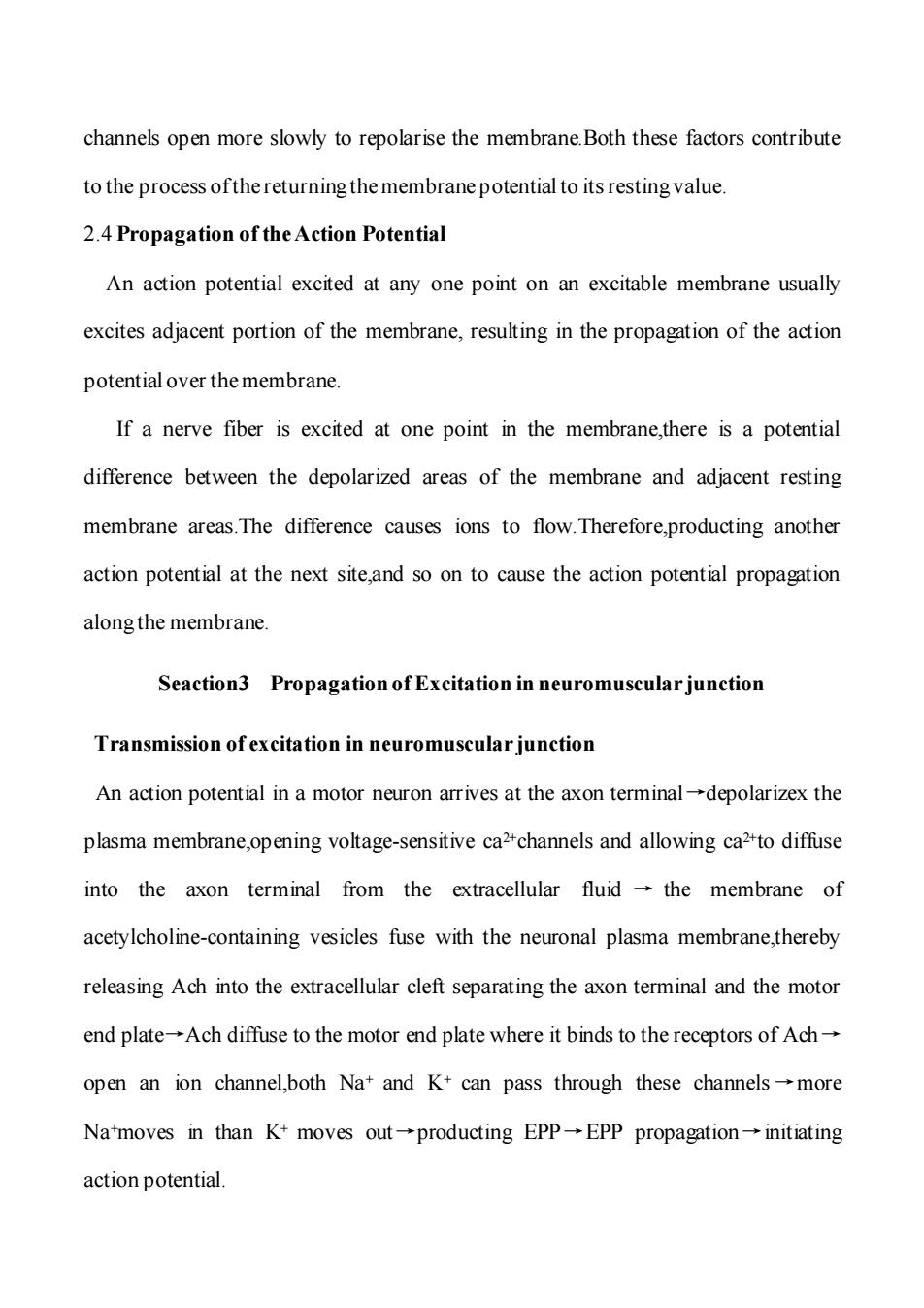
channels open more slowly to repolarise the membrane.Both these factors contribute to the process ofthe returning the membrane potential to its resting value. 2.4 Propagation of the Action Potential An action potential excited at any one point on an excitable membrane usually excites adjacent portion of the membrane,resulting in the propagation of the action potential over the membrane. If a nerve fiber is excited at one point in the membrane,there is a potential difference between the depolarized areas of the membrane and adjacent resting membrane areas.The difference causes ions to flow.Therefore,producting another action potential at the next site,and so on to cause the action potential propagation along the membrane. Seaction3 Propagation of Excitation in neuromuscular junction Transmission of excitation in neuromuscular junction An action potential in a motor neuron arrives at the axon terminal-depolarizex the plasma membrane,opening voltage-sensitive ca2+channels and allowing ca2-to diffuse into the axon terminal from the extracellular fluid -the membrane of acetylcholine-containing vesicles fuse with the neuronal plasma membrane,thereby releasing Ach into the extracellular cleft separating the axon terminal and the motor end plate-Ach diffuse to the motor end plate where it binds to the receptors of Ach- open an ion channel,both Na+and K+can pass through these channels-more Na'moves in than K+moves out-producting EPP-EPP propagation-initiating action potential
channels open more slowly to repolarise the membrane.Both these factors contribute to the process of the returning the membrane potential to its resting value. 2.4 Propagation of the Action Potential An action potential excited at any one point on an excitable membrane usually excites adjacent portion of the membrane, resulting in the propagation of the action potential over the membrane. If a nerve fiber is excited at one point in the membrane,there is a potential difference between the depolarized areas of the membrane and adjacent resting membrane areas.The difference causes ions to flow.Therefore,producting another action potential at the next site,and so on to cause the action potential propagation along the membrane. Seaction3 Propagation of Excitation in neuromuscular junction Transmission of excitation in neuromuscular junction An action potential in a motor neuron arrives at the axon terminal→depolarizex the plasma membrane,opening voltage-sensitive ca2+channels and allowing ca2+to diffuse into the axon terminal from the extracellular fluid → the membrane of acetylcholine-containing vesicles fuse with the neuronal plasma membrane,thereby releasing Ach into the extracellular cleft separating the axon terminal and the motor end plate→Ach diffuse to the motor end plate where it binds to the receptors of Ach→ open an ion channel,both Na+ and K+ can pass through these channels →more Na+moves in than K+ moves out→producting EPP→EPP propagation→initiating action potential
