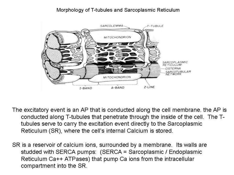
Morphology of T-tubules and Sarcoplasmic Reticulum SARCOLEMMA- T-TUBULE MITOCHONDRION RETTOLUMMIC CISTERNA NERCORBULAR MITOCHONDRION I-BAND A-BAND Z-LINE The excitatory event is an AP that is conducted along the cell membrane.the AP is conducted along T-tubules that penetrate through the inside of the cell.The T- tubules serve to carry the excitation event directly to the Sarcoplasmic Reticulum(SR),where the cell's internal Calcium is stored. SR is a reservoir of calcium ions,surrounded by a membrane.Its walls are studded with SERCA pumps:(SERCA Sarcoplasmic Endoplasmic Reticulum Ca++ATPases)that pump Ca ions from the intracellular compartment into the SR

Schematic of the events involved in excitation-contraction coupling. A L-type Ca channel 3Na* A:Excitation-Ca Release. NCX (a)Calcium enters the cell via L-type Ca channels situated in the surface membrane and (as shown) Ca2 SR in transverse tubules. RyR SERCA (b)Some of this Ca binds to the ryanodine receptor (RyR)making it open and thereby releasing much phospholamban more Ca in a process known as calcium-induced calcium release. (c)Ca release results in the systolic Ca transient and thence contraction. B 0.8r (d)Relaxation occurs by Ca being reduced to resting Ca transient levels by a combination of uptake into the amplitude sarcoplasmic reticulum(SR)via SERCA and (umol/L) removal from the cell by the electrogenic Na-Ca exchange(NCX). 0.0 (B)The steep dependence of the amplitude of the 0 25 50 systolic Ca transient on SR Ca content. SR Ca content(umol/L)

Excitation:AP conducted down the T-Tubule Excitation Ca2+ 2 Ca2+ Sarcotubule Ca2+ Cisterna troponin C 1-L-type Ca2+channel 2-Ca2+-gated Ca2+channel(ryanodine receptor) Notable exceptions to this scheme: Atrial myocytes:Do not have T-tubules;Calcium events have to move into the cell interior by diffusion and are thus slow.As a result they are relatively slow contracting. Purkinje fibers.Do not have T-tubules.These are cells that are specialized for conduction,not contraction.They are more like an axon than a muscle cell.They are minimally contractile

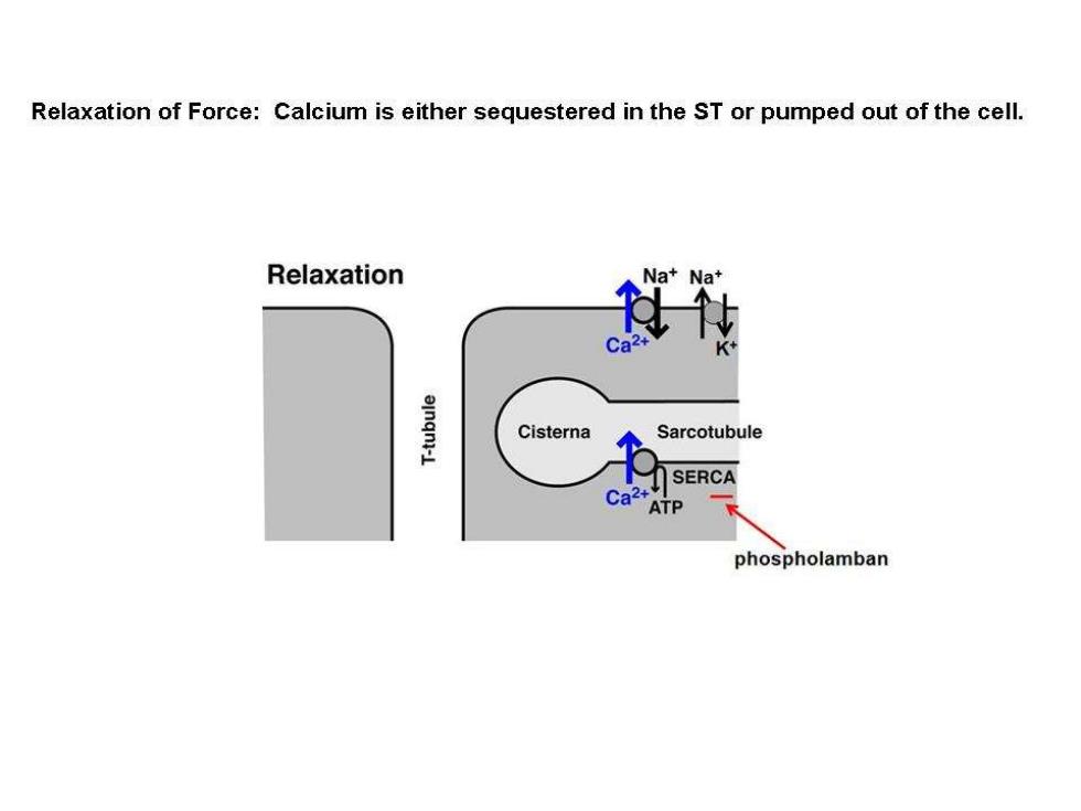
Relaxation of Force:Calcium is either sequestered in the ST or pumped out of the cell. Relaxation Na+Na+ Ca einqn1-L Cisterna Sarcotubule SERCA phospholamban

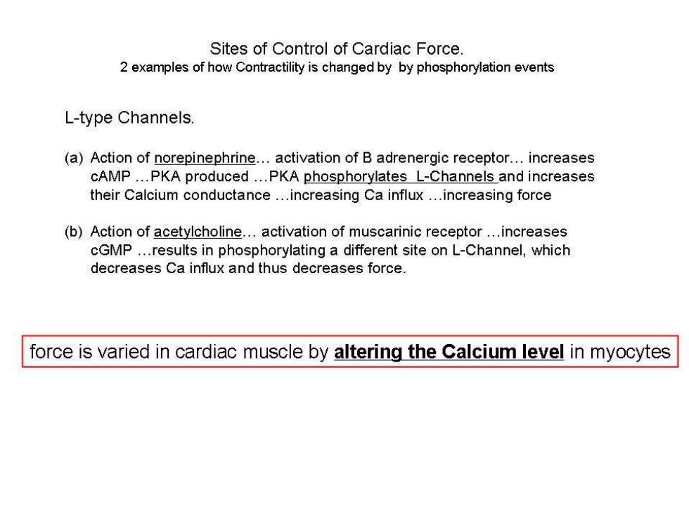
Sites of Control of Cardiac Force. 2 examples of how Contractility is changed by by phosphorylation events L-type Channels. (a)Action of norepinephrine...activation of B adrenergic receptor...increases cAMP...PKA produced...PKA phosphorylates L-Channels and increases their Calcium conductance...increasing Ca influx...increasing force (b)Action of acetylcholine...activation of muscarinic receptor...increases cGMP...results in phosphorylating a different site on L-Channel,which decreases Ca influx and thus decreases force. force is varied in cardiac muscle by altering the Calcium level in myocytes

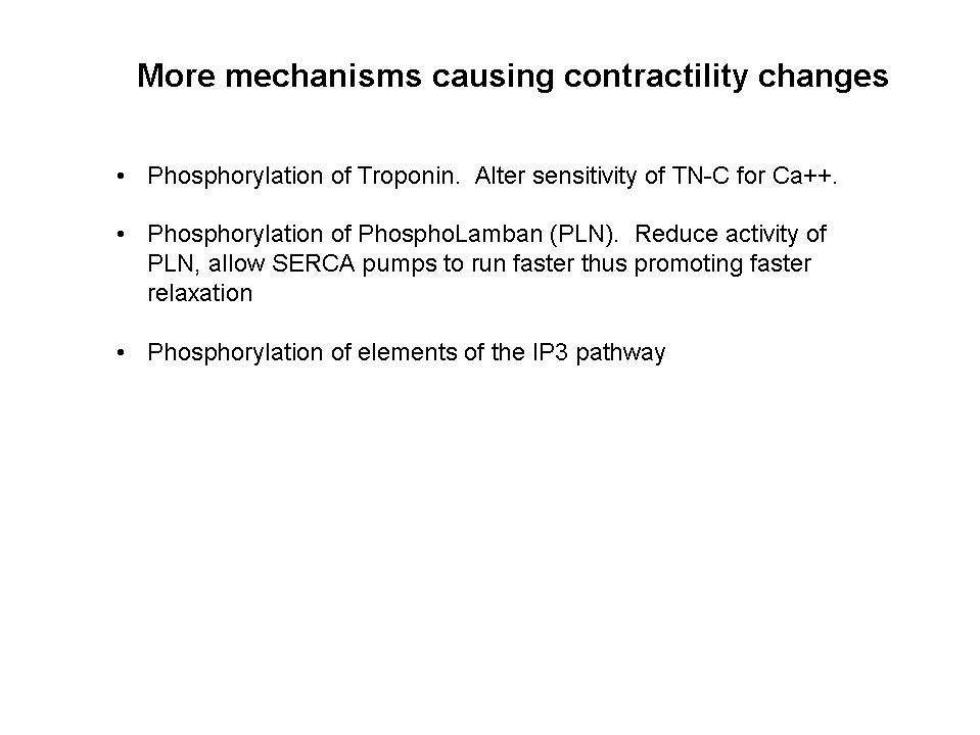
More mechanisms causing contractility changes Phosphorylation of Troponin.Alter sensitivity of TN-C for Ca++. Phosphorylation of PhosphoLamban(PLN).Reduce activity of PLN,allow SERCA pumps to run faster thus promoting faster relaxation Phosphorylation of elements of the IP3 pathway

Ventricular Muscle Excitation-Contraction Timing [Ca] AP 】 Contraction 200ms