p.191 in the textbook X-ray microanalysis Lanting Zhang School of Materials Science and Engineering SJTU lantingzh@sjtu.edu.cn 1
X-ray microanalysis Lanting Zhang School of Materials Science and Engineering, SJTU lantingzh@sjtu.edu.cn 1 p.191 in the textbook
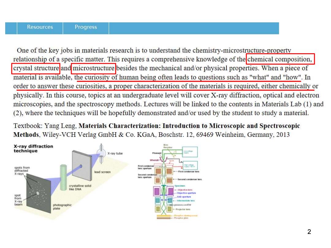
Resources Progress One of the key jobs in materials research is to understand the chemistry-microstructure-property relationship of a specific matter.This requires a comprehensive knowledge of thechemical composition, crystal structure and microstructurebesides the mechanical and/or physical properties.When a piece of material is available,the curiosity of human being often leads to questions such as "what"and "how".In order to answer these curiosities,a proper characterization of the materials is required,either chemically or physically.In this course,topics at an undergraduate level will cover X-ray diffraction,optical and electron microscopies,and the spectroscopy methods.Lectures will be linked to the contents in Materials Lab (1)and (2),where the techniques will be hopefully demonstrated and/or used by the student to study a material. Textbook:Yang Leng,Materials Characterization:Introduction to Microscopic and Spectroscopic Methods,Wiley-VCH Verlag GmbH Co.KGaA,Boschstr.12,69469 Weinheim,Germany,2013 X-ray diffraction technique x-ray tube spots from x-rays ead screen crystalline solid lke DNA spot from X-ray beam photographic 2
2
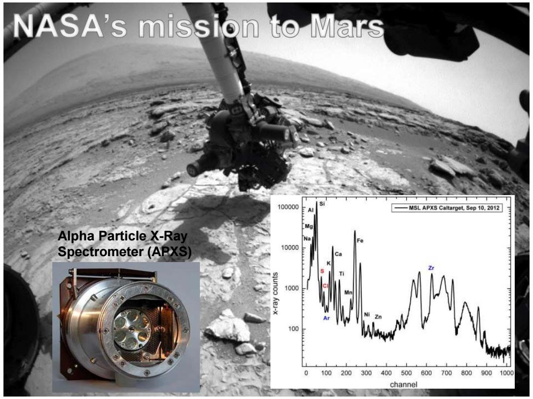
NASA's mission to Mars 100000 MSL APXS Caltarget,Sep 10,2012 Mg Alpha Particle X-Ray Na Fe Spectrometer (APXS 10000 Ca K Zr 1000 Ar 100 0 100200300 400500600700800900 1000 channel
3 Alpha Particle X-Ray Spectrometer (APXS)
How to characterize chemical composition? ·Classical methods -Wet chemistry:turn into a solution Spectroscopy methods -XRF,FTIR,XPS,EDS,SIMS,ICP-MS,WDS. EELS etc -Spatial resolution of the methods?- 4
How to characterize chemical composition? • Classical methods – Wet chemistry: turn into a solution • Spectroscopy methods – XRF, FTIR, XPS, EDS, SIMS, ICP-MS, WDS, EELS etc • Spatial resolution of the methods? 4

Important signals in analytical electron microscopy High energy particle X-ray Incident high kV beam Electron etc of electrons Backscattered e Secondary e (SE) Characteristic (BSE) x-rays Auger e Visible light Bulk(SEM) Absorbed e← e-hole pairs Foil (TEM) Bremsstrahlung X-rays (noise) Elastically Inelastically Interaction scattered e- scattered e- Volume Direct (transmitted) beam Characteristic X-rays constitute a fingerprint of the local chemistry. 5
Important signals in analytical electron microscopy Characteristic X 5 -rays constitute a fingerprint of the local chemistry. High energy particle X-ray Electron etc
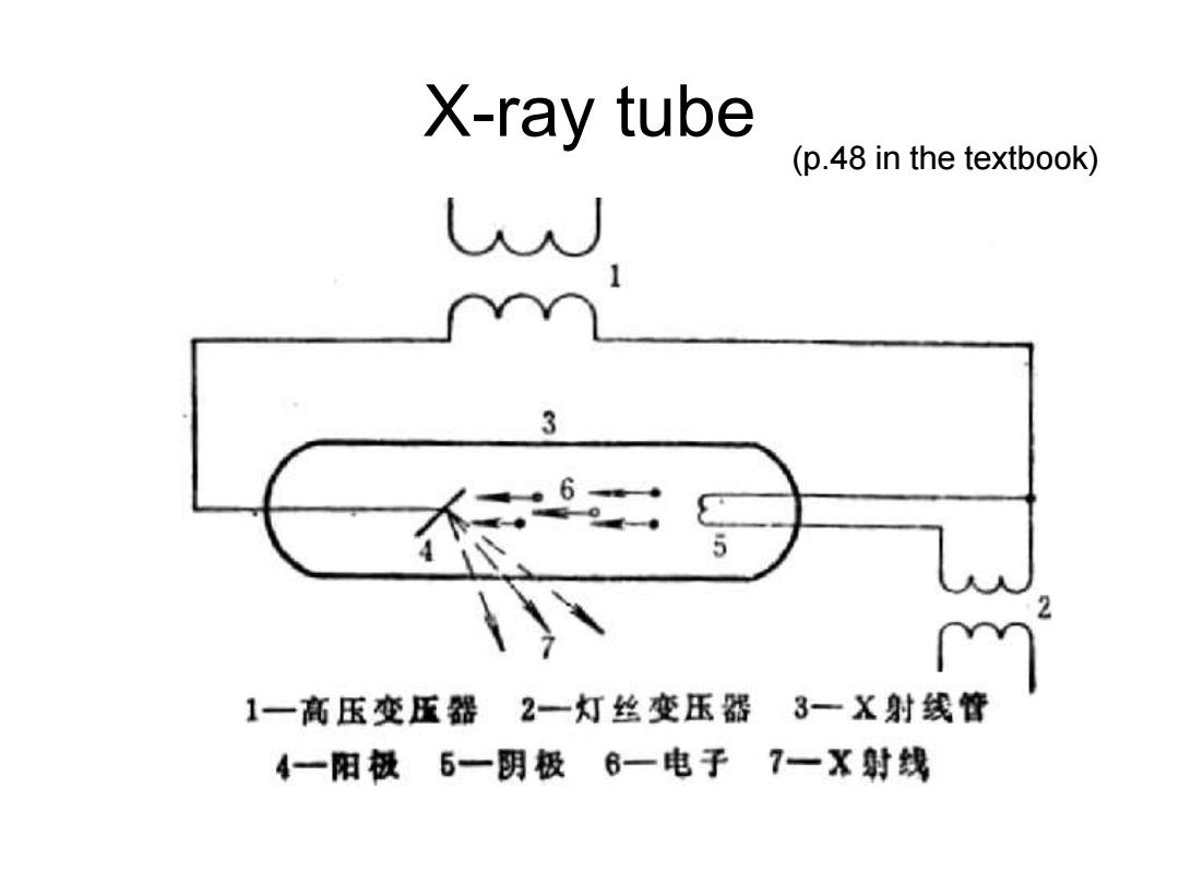
X-ray tube (p.48 in the textbook) 3 5 1一高压变压器2一灯丝变压器 3一X射线管 4一阳极5一阴极6一电子7一X射线
X-ray tube (p.48 in the textbook)
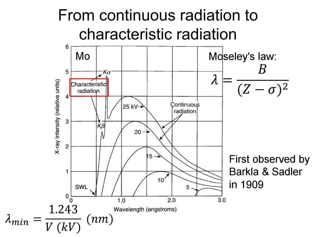
From continuous radiation to characteristic radiation 6 Mo Moseley's law: Ka B Characteristid λ= radiation (Z-0)2 25kV> Continuous radiation 3 网 20 2 15 First observed by 1 Barkla Sadler 10 SWL 5、 in1909 1.0 2.0 3.0 1.243 Wavelength(angstroms) 入mim V(kV) (nm)
From continuous radiation to characteristic radiation Mo First observed by Barkla & Sadler in 1909 Moseley's law: 𝜆 = 𝐵 (𝑍 − 𝜎) 2 𝜆𝑚𝑖𝑛 = 1.243 𝑉 (𝑘𝑉) (𝑛𝑚)
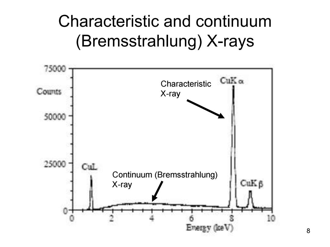
Characteristic and continuum (Bremsstrahlung)X-rays 75000 Characteristic CuKa Counts X-ray 50000 25000- CuL Continuum(Bremsstrahlung) X-ray CuKB 10 Energy (eV) 8
Characteristic and continuum (Bremsstrahlung) X-rays 8 Characteristic X-ray Continuum (Bremsstrahlung) X-ray
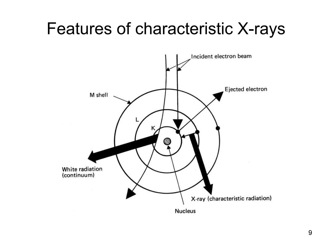
Features of characteristic X-rays Incident electron beam Ejected electron M shell White radiation (continuum) X-ray (characteristic radiation) Nucleus 9
Features of characteristic X-rays 9
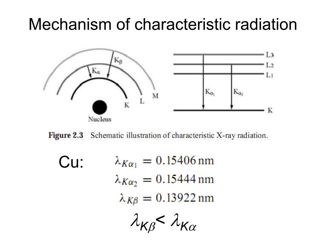
Mechanism of characteristic radiation L3 Kp L2 LI M K K Nucleus Figure 2.3 Schematic illustration of characteristic X-ray radiation Cu: 入Ka1=0.15406nm 入Ka2=0.15444nm 入K6=0.13922nm KB九Ka
Cu: K< K Mechanism of characteristic radiation